Summary
Revista Brasileira de Ginecologia e Obstetrícia. 2012;34(12):531-535
Summary
Revista Brasileira de Ginecologia e Obstetrícia. 2012;34(12):536-543
DOI 10.1590/S0100-72032012001200002
PURPOSES: To identify and to analyze maternal mortality causes, according to hospital complexity levels. METHODS: A descriptive-quantitative cross-sectional study of maternal deaths that occurred in hospitals in Paraná, Brazil, during the periods from 2005 to 2007 and from 2008 to 2010. Data from case studies of maternal mortality, obtained by the State Committee for Maternal Mortality Prevention, were utilized. The study focused on variables such as site and causes of death, hospital transfer, and avoidability. Maternal mortality rate, proportions, and hospital lethality ratio were calculated according to subgroups of low and high-risk pregnancy reference hospitals. RESULTS: Maternal mortality rate, including late maternal deaths, was 65.9 per 100.000 live-borns (from 2008 to 2010). Almost 90% of all maternal deaths occurred in the hospital environment, in both periods. The hospital lethality ratio at the high-risk pregnancy reference hospital was 158.4 deaths per 100,000 deliveries during the first period and 132.5/100,000 during the second, and the main causes were pre-eclampsia/eclampsia, puerperal infection, urinary tract infection, and indirect causes. At the low-risk pregnancy reference hospitals, the hospital lethality ratios were 76.2/100,000 and 80.0/100,000, and the main causes of death were hemorrhage, embolism, and anesthesia complications. In 64 (2005 - 2007) and in 71% (2008 - 2010) of the cases, the patients died in the same hospital of admission. During the second period, 90% of the casualties were avoidable. CONCLUSIONS: Hospitals of both levels of complexity are having difficulties in treating obstetric complications. Professional training for obstetric emergency assistance and the monitoring of protocols at all hospital levels should be considered by the managers as a priority strategy to reduce avoidable maternal deaths.
Summary
Revista Brasileira de Ginecologia e Obstetrícia. 2012;34(12):544-549
DOI 10.1590/S0100-72032012001200003
PURPOSE: To describe the epidemiological cases and microbiological profile of Streptococcus agalactiae serotypes isolated from infected newborns of a Women's Health Reference Centre of Campinas, São Paulo, Brazil. METHODS: Cross-sectional laboratory survey conducted from January 2007 to December 2011. The newborns' strains, isolated from blood and cerebrospinal fluid samples, were screened by hemolysis on blood ágar plates, Gram stain, catalase test, CAMP test, hippurate hydrolysis or by microbiological automation: Vitek 2 BioMerieux®. They were typed by PCR, successively using specific primers for species and nine serotypes of S. agalactiae. RESULTS: Seven blood samples, one cerebrospinal fluid sample and an ocular sample, were isolated from nine newborns with infections caused by S. agalactiae, including seven cases of early onset and two of late onset. Only one of these cases was positive for paired mother-child samples. Considering that 13,749 deliveries were performed during the study period, the incidence was 0.5 cases of GBS infections of early onset per 1 thousand live births (or 0.6 per 1 thousand, including two cases of late onset) with 1, 3, 2, zero and 3 cases (one early and two late onset cases), respectively, for the years from 2007 to 2011. It was possible to apply PCR to seven of nine samples, two each of serotypes Ia and V and three of serotype III, one from a newborn and the other two from a paired mother-child sample. CONCLUSIONS: Although the sample was limited, the serotypes found are the most prevalent in the literature, but different from the other few Brazilian studies available, except for type Ia.
Summary
Revista Brasileira de Ginecologia e Obstetrícia. 2012;34(12):563-567
DOI 10.1590/S0100-72032012001200006
PURPOSE: To investigate the relationship between periodontitis and osteoporosis, using a case-control study about periodontal status of postmenopausal women. METHODS: A total of 99 postmenopausal women were divided into three groups: normal bone (Gn, n=45), osteopenia (Gpenia, n=31) and osteoporosis (Gporosis, n=23). The categorization of bone mass was measured by dual energy absorptiometry with X-rays in the lumbar spine (L2 - L4), by assessing bone mineral density. Clinical attachment level (CAL), gingival bleeding index (GI), plaque index (PI), and probing depth (PD) were determined in all participants by a single examiner. The data were submitted to BioEstat 2.0 software through parametric analysis of variance (ANOVA) and the Bonferroni test, with the level of significance set at 5%. RESULTS: Women with osteoporosis presented the highest percentage of periodontal disease, with higher average CAL (2.6±0.4 mm) and PD (2.8±0.6 mm), GI (72.8±25.9 mm) and PI (72.9±24.2 mm). Statistical analysis revealed a significant difference in periodontal situation between Gn and Gporosis (p=0,01) and between Gpenia and Gporosis (p=0,03). CONCLUSION: Osteoporosis may have an influence on periodontal condition, based on the relation between periodontitis and osteoporosis in postmenopausal women.
Summary
Revista Brasileira de Ginecologia e Obstetrícia. 2012;34(12):568-574
DOI 10.1590/S0100-72032012001200007
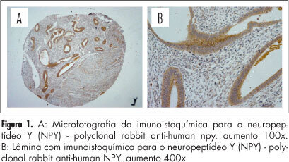
PURPOSE: To evaluate the expression of neurotrophic (NGF, NPY and VIP) and pro-inflammatory (TNF-α) mediators in the rectum and sigmoid fragments compromised by endometriosis. METHODS: Twenty-four patients were selected to undergo surgical treatment of endometriosis of the rectum and sigmoid colon with a segmental resection technique, followed by end-to-end anastomosis with a circular stapler from January 2005 to December 2007. The study included premenopausal women who underwent surgical treatment for deep endometriosis infiltrating the rectum with involvement of the rectum and sigmoid, reaching the level of the muscle layer, submucosa or mucosa. Twenty-four rectum and sigmoid fragments with histologically confirmed endometriosis, one from each of the 24 selected patients, were used for the study group. For the control group, we used a fragment of the distal resection margin called anastomosis ring from each of the 24 patients enrolled in the study. Samples were grouped into Tissue Micro Array (TMA) blocks and subjected to immunohistochemistry to evaluate the expression of tumor necrosis factor alpha (TNF-α), nerve growth factor (NGF), neuropeptide Y (NPY) and P vasoactive intestinal peptide (VIP), followed by semiquantitative analysis of immunostaining by reading the relative optical density (OD). RESULTS: There was higher optical density relative to TNF-α immunostaining and NGF in the study group (samples with intestinal endometriosis), DO=0.01, for the two proteins, respectively (p<0.05), compared to controls without endometriosis. There was no statistically significant difference in the optical density of immunostaining of NPY and VIP. CONCLUSION: We identified increased immunostaining of TNF-α antibodies and fragments of NGF in the rectum and sigmoid compromised by endometriosis compared to disease-free controls. We did not identify any statistical difference in immunostaining of NPY and VIP proteins.

Summary
Revista Brasileira de Ginecologia e Obstetrícia. 2012;34(12):575-581
DOI 10.1590/S0100-72032012001200008
PURPOSE: To compare serum anti-Mullerian hormone (AMH) levels on the seventh day of ovarian stimulation between normal and poor responders. METHODS: Nineteen women aged ≥35, presenting with regular menses, submitted to ovarian stimulation for assisted reproduction were included. Women with endometriosis, polycystic ovarian syndrome or those who were previously submitted to ovarian surgery were excluded. On the basal and seventh day of ovarian stimulation, a peripheral blood sample was drawn for the determination of AMH, FSH and estradiol levels. AMH levels were assessed by ELISA and FSH, and estradiol by immunochemiluminescence. At the end of the stimulation cycle patients were classified as normal responders (if four or more oocytes were obtained during oocyte retrieval) or poor responders (if less than four oocytes were obtained during oocyte retrieval or if the cycle was cancelled due to failure of ovulation induction) and comparatively analyzed by the t-test for hormonal levels, length of ovarian stimulation, number of follicles retrieved, and number of produced and transferred embryos. The association between AMH and these parameters was also analyzed by the Spearman correlation test. RESULTS: There was no significant difference between groups for basal or the seventh day as to AMH, FSH and estradiol levels. There was a significant correlation between seventh day AMH levels and the total amount of exogenous FSH used (p=0.02). CONCLUSIONS: AMH levels on the seventh day of the ovarian stimulation cycle do not seem to predict the pattern of ovarian response and their determination is not recommended for this purpose.