Summary
Revista Brasileira de Ginecologia e Obstetrícia. 2002;24(3):161-166
DOI 10.1590/S0100-72032002000300003
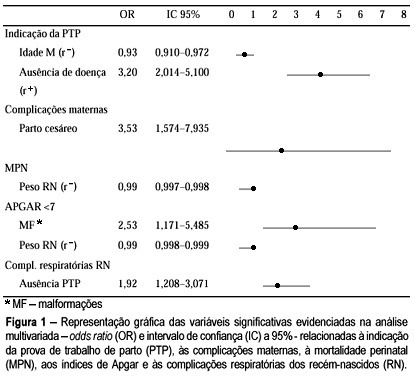
Purpose: to study trial of labor (TOL) for vaginal birth after one previous cesarean section. Methods: this is a retrospective cohort study that included 438 pregnant women with one previous cesarean section and their 450 newborns. They were divided into two groups - with and without TOL. The minimum sample size was 121 pregnant mothers per group. TOL was considered as an independent variable and vaginal birth and maternal and perinatal complication frequency as dependent variables. Both univariate and multivariate analyses were performed. The comparison of observed frequencies (%) was analyzed by the chi-squared test (chi²) with 5% significance, and linear regression from the odds ratio (OR) and confidence interval of 95% (CI95%). Results: TOL was used in 59.2% of vaginal deliveries. It was less used in women over 40 years (2.7% vs 6.7%) and in those with clinical or obstetrical diseases such as arterial hypertension (7.0%) and bleeding in the third trimester (0.3%). There was a higher risk for puerperal complications with cesarean deliveries (OR = 3.53, CI 95% = 1.57-7.93), independent of TOL. Perinatal mortality was dependent on neonatal weight and fetal malformations, not on TOL. Newborns from mothers not submitted to TOL were at a higher risk for developing breathing complications (OR = 1.92 CI 95% = 1.20-3.07). Conclusions: The results confirm that trial of labor after a previous cesarean section is a safe method - assisting vaginal delivery in 59.2% of births and not interfering with maternal and perinatal mortality. It is a treatment that should be stimulated.

Summary
Revista Brasileira de Ginecologia e Obstetrícia. 2002;24(3):195-199
DOI 10.1590/S0100-72032002000300008
Purpose: to evaluate, in a prospective way, the importance of ultrasound features of solid breast lesions in the differentiation between benign and malignant lumps. Methods: one hundred and forty-two patients with solid breast lesions, from the Department of Gynecology and Obstetrics of the Federal University of Goias (Brazil), were included in the trial. All ultrasound examinations were performed by a training doctor, always supervised by an experienced professional. The characteristics of the lesions studied were: shape, retrotumoral echoes, internal echoes, oriented diameter, halo of bright echoes and Cooper ligaments. Each of the ultrasound features was compared to the results of the histological examination. Results: among the 142 patients included in the trial, 90 (63%) had their lesions excised, and 77 (86%) had pathologic diagnoses of benign tumors and 13 (14%) of malignant tumors. The following characteristics were statistically significant in the diagnosis of the breast cancer (c²): masses with retrotumoral shadowing (p=0.0001), irregular shape (p=0.0007), heterogeneous internal echoes (p=0.0015) and vertically oriented - taller than wide (p<0.0001). The presence of halo of bright echoes anterior to the lump and the presence of wider Cooper ligaments were not related to the correct diagnosis of malignancy in this trial. Conclusion: ultrasound is a diagnostic method that can help physicians between the differentiation of benign and malignant breast lumps. The presence of retrotumoral shadowing, irregular shape, heterogeneous internal echoes and vertical orientation - lesions taller than wide - were related to the pathologic diagnosis of breast malignancies.
Summary
Revista Brasileira de Ginecologia e Obstetrícia. 2002;24(3):187-192
DOI 10.1590/S0100-72032002000300007
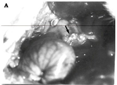
Purpose: in order to maintain the gonadal function after oophorectomy, morphofunctional aspects of ovarian autotransplantation to the greater omentum and the best kind of implantation, intact or sliced, were investigated. Methods: forty cycling female Wistar rats were randomly divided into four groups: Group I (n = 5), control - laparotomy; Group II (n = 5), bilateral oophorectomy; Group III (n = 10), intact ovarian autotransplantation to the greater omentum; and Group IV (n = 10), sliced ovarian autotransplantation, both to the greater omentum. The estrous cycle was investigated in the third and sixth postoperative months and histological studies of the ovarian implants were carried out considering: degeneration, fibrosis, inflammatory reaction, angiogenesis, follicular cysts, follicular development and corpora lutea. Results: the animals of Group I preserved the cycling sequence. The rats of Group II remained in diestrus. In Group III, 11 rats remained in diestrus, three presented incomplete cycles and one showed normal cycle. In Group IV, three animals remained in diestrus, eight showed incomplete cycles and four showed normal cycles. The histology of the ovaries of Group III was normal in ten female rats; however, the ovaries of the other five animals presented degeneration. In Group IV, 14 female rats had ovaries with preserved morphological aspect, and signs of degeneration occurred in one. Conclusions: the ovarian autotransplantation to the greater omentum is viable and the sliced form presented better morphofunctional aspects than the intact implants.

Summary
Revista Brasileira de Ginecologia e Obstetrícia. 2002;24(3):181-185
DOI 10.1590/S0100-72032002000300006
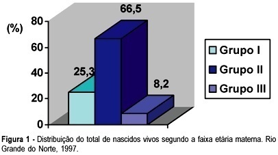
Purpose: to investigate the interactions between maternal age and adverse perinatal outcomes in the State of Rio Grande do Norte. Methods: we analyzed official records of 57,088 infants in the State of Rio Grande do Norte, from January 1997 to December 1997. Data were obtained from the Information System of the Health Ministry, Brazil. The sample was divided into three Groups I, II and III according to maternal age range: 10 to 19 years, 20 to 34, and 35 or more, respectively. The main outcome variables were: length of pregnancy, birth weight and mode of delivery. Statistical analysis was performed using chi² test. Results: preterm deliveries were 4.3% in the adolescent group vs 3.7% in Group II (p = 0.0028). The incidence of cesarean section was higher in Group II than in the other Groups (p<0.001). Low birth weight was significantly higher in Groups I (8.4%) and III (8.3%) when compared with Group II (6.5%) (p<0.0001). Conclusions: we found a higher incidence of lower birth weight and preterm delivery in the adolescent group. In women ³35 years old there was a high incidence of low birth weight and macrosomia. Results suggest that cesarean sections are more common in women aged 20-34 years than in adolescent and older mothers.

Summary
Revista Brasileira de Ginecologia e Obstetrícia. 2002;24(3):175-179
DOI 10.1590/S0100-72032002000300005
Purpose: to determine the presence of asymptomatic amniotic fluid infection in pregnant women, to identify the bacterial agents involved in the infection and to determine the antimicrobial susceptibility in vitro. Methods: amniotic fluid samples were obtained by amniocentesis from 81 pregnant women without labor signs and without suspucion of clinical infection, attended at Maternidade Escola Assis Chateaubriand from August 1997 to January 1999. The presence of aerobic bacteria, strict/facultative anaerobic bacteria and genital mycoplasmas was investigated. The anaerobic bacteria were identified by the ATB SystemÒ (Biolab Mérieux) and mycoplasmas by the IST MycoplasmaÒ kit (Biolab-Mérieux). Results: among the obtained samples, eight (9.8%) showed positive culture and in two samples two different strains were identified. The isolated pathogens were Ureaplasma urealyticum (7 cases, 8.6%), Mycoplasma hominis (1 case, 1.2%) and Peptostreptococcus sp (2 cases, 2.4%). The antimicrobial susceptibility was characterized by great mycoplasma resistance to erythromycin (37.5%) and no resistance to cyclins. Conclusions: the percentage of asymptomatic infections was high, and furthe research is necessary to evaluate the asymptomatic infection consequences in pregnant women and their newborns, involving methods that identify genital mycoplasmas, which were the most frequently isolated bacteria.
Summary
Revista Brasileira de Ginecologia e Obstetrícia. 2002;24(1):37-43
DOI 10.1590/S0100-72032002000100006
Purpose: to determine the factors associated with the detection of a microinvasive carcinoma in the cervical cone of women with a previous colposcopically directed biopsy compatible with cervical intraepithelial neoplasia (CIN) 3 and to evaluate the proportion of involved margins. Patients and methods: we reviewed the medical records of 385 women (mean age: 39 years) submitted to cold conization or conization by high frequency surgery (HFS) with a loop during the period from January 1993 to July 2000. These procedures were indicated on the basis of a biopsy compatible with (CIN) 3. Results: the diagnosis of the cone was compatible with (CIN) 3 in 243 (63%) women and with (CIN) 2 in 13 (3%). Only 10 presented HPV/CIN 1 (3%) and eight had no residual disease in the cone. However, 101 (26%) women presented a microinvasive carcinoma in the cone and 10 (3%) presented a frankly invasive carcinoma. Age, menstrual status and number of deliveries were not related to the severity of the cone lesion. Women with oncologic colpocytology changes suggestive of invasion presented a significantly higher risk of having a microinvasive or invasive carcinoma as determined by final histology (p<0.01), although 52 of the 243 women with CIN 2 or CIN 3 in the cone also showed a suggestion of invasion at colpocytology. Among the women with CIN 2 or 3, the epithelium was white in 44%, dotted in 21%, and mosaic-like in 17%. This proportion was similar for women with a microinvasive or invasive carcinoma, with these images being detected in 37%, 23% and 21% of the cases, respectively. Involvement of the cone margins was significantly higher among women submitted to HFS (49%) than among those submitted to cold conization (29%). Conclusion: the absence of independent clinical and colposcopic factors associated with the detection of a microinvasive carcinoma in women submitted to conization on the basis of a biopsy compatible with (CIN) 3 justifies the conical excision of the squamocolumnar junction in high grade cervical lesions.
Summary
Revista Brasileira de Ginecologia e Obstetrícia. 2002;24(1):51-57
DOI 10.1590/S0100-72032002000100008
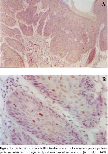
Purpose: to evaluate p53 overexpression value in vulvar intraepithelial neoplasia (VIN) III recurrence/progression. Methods: twenty patients with undifferentiated VIN III were selected and followed up every six months for four years and divided into two groups: fourteen without and six with recurrence/progression lesion. The recurrence/progression cases were distributed as follows: in three patients recurrence occurred only once; in two, twice, and only one progressed to squamous cancer. In both groups the site of vulvar lesion and p53 overexpression and immunostaining pattern were analyzed. A similar study was performed in recurrence/progression cases, besides the analysis of the time interval to occur the arise of recurrence/progression. Results: recurrence was observed in 25% of the cases and, in 5%, progression to carcinoma. The mean time interval for recurrence was 24.5 months. Multifocal location of the initial lesion was the predominant form (50%) in both groups. In the majority of the cases (87.5%) recurrence/progression occurred at the same site of the initial vulvar lesion. p53 overexpression was observed in 50% of the VIN III primary lesions and in 75% of the recurrence/progression cases. Conclusions: p53 overexpression seems to play an important role in VIN III pathogenesis and may predict the clinical course of the lesions. VIN III recurrence/progression has a tendency to occur in the same area of the initial lesion, suggesting the presence of molecular disturbance.
