Summary
Revista Brasileira de Ginecologia e Obstetrícia. 2019;41(4):220-229
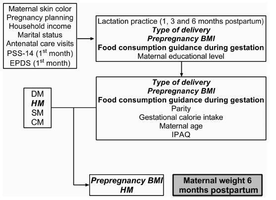
Different intrauterine environments may influence the maternal prepregnancy body weight (BW) variation up to 6 months postpartum. The objective of the present study was to verify the association of sociodemographic, obstetric, nutritional, and behavioral factors with weight variation in women divided into four groups: hypertensive (HM), diabetic (DM), smokers (SM), and control mothers (CM).
It was a convenience sample of 124 postpartum women recruited from 3 public hospitals in the city of Porto Alegre, state of Rio Grande do Sul, Brazil, between 2011 and 2016.Multiple linear regressions and generalized estimating equations (GEE) were conducted to identify the factors associated with maternal weight variation. For all GEE, the maternal weight measurements were adjusted for maternal height, parity, educational level, and the type of delivery, and 3 weight measurements (prepregnancy, preceding delivery, and 15 days postpartum) were fixed.
A hierarchical model closely associated the maternal diagnosis of hypertension and a prepregnancy body mass index (BMI) classified as overweight with maternal weight gain measured up to the 6th month postpartum (the difference between the maternal weight at 6months postpartum and the prepregnancy weight). These results showed that the BW of the HM group and of overweight women increased ~ 5.2 kg 6 months postpartum, compared with the other groups. Additionally, women classified as overweight had a greater BW variation of 3.150 kg.
This evidence supports the need for specific nutritional guidelines for gestational hypertensive disorders, as well as great public attention for overweight women in the fertile age.

Summary
Revista Brasileira de Ginecologia e Obstetrícia. 2019;41(1):24-30
The aim of this study is to evaluate whether exposure to different environmental lighting conditions affects the reproductive parameters of pregnant mice and the development of their offspring.
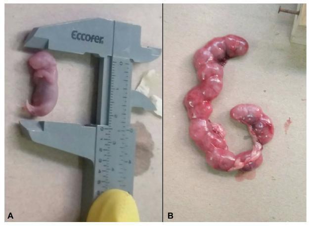
Fifteen pregnant albino mice were divided into three groups: light/dark, light, and dark. The animalswere euthanized on day 18 of pregnancy following the Brazilian Good Practice Guide for Euthanasia of Animals.Maternal and fetal specimens weremeasured and collected for histological evaluation. Analysis of variance (ANOVA) test was used for comparison of the groups considering p ≤ 0.05 to be statistically significant.
There was no significant difference in the maternal variables between the three groups. Regarding fetal variables, significant differences were observed in the anthropometric measures between the groups exposed to different environmental lighting conditions, with the highest mean values in the light group. The histological evaluation showed the same structural pattern of the placenta in all groups, which was within the normal range. However, evaluation of the uterus revealed a discrete to moderate number of endometrial glands in the light/dark and light groups, which were poorly developed in most animals. In the fetuses, pulmonary analysis revealed morphological features consistent with the transition from the canalicular to the saccular phase in all groups.
Exposure to different environmental lighting conditions had no influence on the reproductive parameters of female mice, while the offspring of mothers exposed to light for 24 hours exhibited better morphometric features.

Summary
Revista Brasileira de Ginecologia e Obstetrícia. 2019;41(1):31-36
To evaluate the rs42524 polymorphism of the procollagen type I alpha (α) 2 (COL1A2) gene as a factor related to the development of pelvic organ prolapse (POP) in Brazilian women.
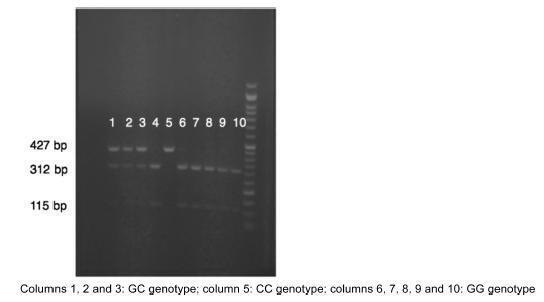
The present study involved 112 women with POP stages III and IV (case group) and 180 women with POP stages zero and I (control group). Other clinical data were obtained by interviewing the patients about their medical history, and blood was also collected from the volunteers for the extraction of genomic DNA. The promoter region of the COL1A2 gene containing the rs42524 polymorphism was amplified, and the discrimination between the G and C variants was performed by digestion of the polymerase chain reaction (PCR) products with the MspA1I enzyme followed by agarose gel electrophoresis analysis.
A total of 292 women were analyzed. In the case group, 71 had the G/G genotype, 33 had the G/C genotype, and 7 had the C/C genotype. In turn, the ratio in the control group was 117 G/G, 51 G/C, and 11 C/C. There were no significant differences between the groups.
Our data did not show an association between the COL1A2 polymorphism and the occurrence of POP.

Summary
Revista Brasileira de Ginecologia e Obstetrícia. 2019;41(1):17-23
To assess and compare the sensitivity and specificity of ultrasonography and magnetic resonance imaging in the diagnosis of placenta accreta in patients with placenta previa.
This retrospective cohort study included 37 women, and was conducted between January 2013 and October 2015; 16 out of the 37 women suffered from placenta accreta. Histopathology was considered the gold standard for the diagnosis of placenta accreta; in its absence, a description of the intraoperative findings was used. The associations among the variables were investigated using the Pearson chi-squared test and the Mann-Whitney U-test.
The mean age of the patients was 31.8 ± 7.3 years, the mean number of pregnancies was 2.8 ± 1.1, the mean number of births was 1.4 ± 0.7, and the mean number of previous cesarean sections was 1.2 ± 0.8. Patients with placenta accreta had a higher frequency of history of cesarean section than those without it (63.6% versus 36.4% respectively; p < 0.001). The mean gestational age at birth among women diagnosed with placenta previa accreta was 35.4 ± 1.1 weeks. The mean birth weight was 2,635.9 ± 374.1 g. The sensitivity of the ultrasound was 87.5%, with a positive predictive value (PPV) of 65.1%, and a negative predictive value (NPV) of 75.0%. The sensitivity of the magnetic resonance imaging was 92.9%, with a PPV of
The ultrasound and the magnetic resonance imaging showed similar sensitivity and specificity for the diagnosis of placenta accreta.
Summary
Revista Brasileira de Ginecologia e Obstetrícia. 2019;41(1):11-16
To evaluate the accuracy of the diagnosis of fetal heart diseases obtained through ultrasound examinations performed during the prenatal period compared with the postnatal evaluation.
A retrospective cohort study with 96 pregnant women who were attended at the Echocardiography Service and whose deliveries occurred at the Complexo Hospitalar Santa Casa de Porto Alegre, in the state of Rio Grande do Sul, Brazil. Risk factor assessment plus sensitivity and specificity analysis were used, comparing the accuracy of the screening for congenital heart disease by means of obstetrical ultrasound and morphological evaluation and fetal echocardiography, considering p < 0.05 as significant. The present study was approved by the Research Ethics Committee of the Institution.
The analysis of risk factors shows that 31.3% of the fetuses with congenital heart disease could be identified by anamnesis. The antepartum echocardiography demonstrated a sensitivity of 97.7%, a specificity of 88.9%, and accuracy of 93% in the diagnosis of congenital heart disease. A sensitivity of 29.3% was found for the obstetric ultrasound, of 54.3% for themorphological ultrasound, and of 97.7% for the fetal echocardiography. The fetal echocardiography detected fetal heart disease in 67.7% of the cases, the morphological ultrasound in 16.7%, and the obstetric ultrasound in 11.5% of the cases.
There is a high proportion of congenital heart disease in pregnancies with no risk factors for this outcome. Faced with the disappointing results of obstetric ultrasound for the detection of congenital heart diseases and the current unfeasibility of universal screening of congenital heart diseases through fetal echocardiography, the importance of the fetal morphological ultrasound and its performance by qualified professionals is reinforced for a more appropriate management of these pregnancies.
Summary
Revista Brasileira de Ginecologia e Obstetrícia. 2019;41(1):04-10
To assess the association between dietary glycemic index (GI) and excess weight in pregnant women in the first trimester of pregnancy.
A cross-sectional study in a sample of 217 pregnant women was conducted at the maternal-fetal outpatient clinic of the Hospital Geral de Fortaleza, Fortaleza, state of Ceará, Brazil, for routine ultrasound examinations in the period between 11 and 13 weeks + 6 days of gestation.Weight and height were measured and the gestational body mass index (BMI) was calculated. The women were questioned about their usual body weight prior to the gestation, considering the prepregnancy weight. The dietary GI and the glycemic load (GL) of their diets were calculated and split into tertiles. Analysis of variance (ANOVA) or Kruskal-Walls and chi-squared (χ2) statistical tests were employed. A crude logistic regression model and a model adjusted for confounding variables known to influence biological outcomes were constructed. A p-value < 0.05 was considered significant for all tests employed.
The sample group presented a high percentage of prepregnancy and gestational overweight (39.7% and 40.1%, respectively). InthetertilewiththehigherGIvalue, therewasa lower dietary intake of total fibers (p = 0.005) and of soluble fibers (p = 0.008). In the third tertile, the dietary GI was associated with overweight in pregnant women in the first trimester of gestation, both in the crude model and in the model adjusted for age, total energy intake, and saturated fatty acids. However, this association was not observed in relation to the GL.
A high dietary GI was associated with excess weight in women in the first trimester of pregnancy.
Summary
Revista Brasileira de Ginecologia e Obstetrícia. 2018;40(11):705-712
To characterize the patterns of cell differentiation, proliferation, and tissue invasion in eutopic and ectopic endometrium of rabbits with induced endometriotic lesions via a well- known experimental model, 4 and 8 weeks after the endometrial implantation procedure.
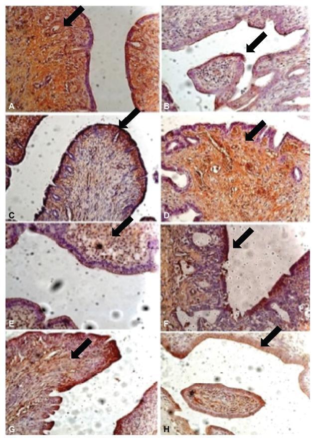
Twenty-nine female New Zealand rabbits underwent laparotomy for endometriosis induction through the resection of one uterine horn, isolation of the endometrium, and fixation of tissue segment to the pelvic peritoneum. Two groups of animals (one with 14 animals, and the other with15) were sacrificed 4 and 8 weeks after endometriosis induction. The lesion was excised along with the opposite uterine horn for endometrial gland and stroma determination. Immunohistochemical reactions were performed in eutopic and ectopic endometrial tissues for analysis of the following markers: metalloprotease (MMP-9) and tissue inhibitor of metalloprotease (TIMP-2), which are involved in the invasive capacity of the endometrial tissue; and metallothionein (MT) and p63, which are involved in cell differentiation and proliferation.
The intensity of the immunostaining for MMP9, TIMP-2, MT, and p63 was higher in ectopic endometria than in eutopic endometria. However, when the ectopic lesions were compared at 4 and 8 weeks, no significant difference was observed, with the exception of the marker p63, which was more evident after 8 weeks of evolution of the ectopic endometrial tissue.
Ectopic endometrial lesions seem to express greater power for cell differentiation and tissue invasion, compared with eutopic endometria, demonstrating a potentially invasive, progressive, and heterogeneous presentation of endometriosis.
