Summary
Revista Brasileira de Ginecologia e Obstetrícia. 2017;39(9):471-479
Using three-dimensional ultrasound (3D-US), we aimed to compare the tape position and the angle formed by the sling arms in different techniques of midurethral sling insertion for the surgical treatment of stress urinary incontinence, three years after surgery. In addition, we examined the correlations between the US findings and the clinical late postoperative results.
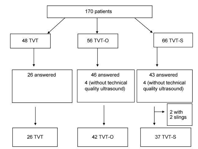
A prospective cross-sectional cohort study of 170 patients who underwent a sling procedure between May 2009 and December 2011 was performed. The final sample, with US images of sufficient quality, included 26 retropubic slings (tension-free vaginal tape, TVT), 42 transobturator slings (tension-free vaginal tape-obturator, TVTO), and 37 single-incision slings (tension-free vaginal tape-Secur, TVT-S). The images (at rest, during the Valsalva maneuver, and during pelvic floor contraction) were analyzed offline by 2 different observers blinded against the surgical and urinary continence status. Group comparisons were performed using the Student t-test, the chi-squared and the Kruskal-Wallis tests, and analyses of variance with Tukey multiple comparisons.
Differences among the groups were found in themean angle of the tape arms (TVT = 119.94°, TVT-O = 141.93°, TVT-S = 121.06°; p < 0.001) and in the distance between the bladder neck and the tape at rest (TVT = 1.65 cm, TVT-O = 1.93 cm, TVTS = 1.95 cm; p = 0.010). The global objective cure rate was of 87.8% (TVT = 88.5%, TVT-O = 90.5%, TVT-S = 83.8%; p = 0.701). The overall subjective cure rate was of 83.8% (TVT = 88.5%, TVT-O = 88.5% and TVT-S = 78.4%; p = 0.514). The slings were located in the mid-urethra in 85.7% of the patients (TVT = 100%, TVT-O = 73.8%, TVTS = 89.2%; p = 0.001), with a more distal location associated with obesity (distal: 66.7% obese; mid-urethra: 34% obese; p = 0.003). Urgency-related symptoms were observed in 23.8% of the patients (TVT = 30.8%, TVT-O = 21.4%, TVT-S = 21.6%; p = 0.630).
The angle formed by the arms of the sling tape was more obtuse for the transobturator slings compared with the angles for the retropubic or single-incision slings. Retropubic slings were more frequently located in the mid-urethra compared with the other slings, regardless of obesity. However, the analyzed sonographic measures did not correlate with the urinary symptoms three years after the surgery.

Summary
Revista Brasileira de Ginecologia e Obstetrícia. 2017;39(9):464-470
To describe the blood flow velocities and impedance indices changes in the uterine arteries of leiomyomatous uteri using Doppler sonography.
This was a prospective, case-control study conducted on 140 premenopausal women with sonographic diagnosis of uterine leiomyoma and 140 premenopausal controls without leiomyomas. Pelvic sonography was performed to diagnose and characterize the leiomyomas. The hemodynamics of the ascending branches of both main uterine arteries was assessed by Doppler interrogation. Statistical analysis was performed mainly using non-parametric tests.
The median uterine volume of the subjects was 556 cm3, while that of the controls was 90.5 cm3 (p < 0.001). The mean peak systolic velocity (PSV), end-diastolic velocity (EDV), time-averaged maximum velocity (TAMX), time-averaged mean velocity (Tmean), acceleration time (AT), acceleration index (AI), diastolic/systolic ratio (DSR), diastolic average ratio (DAR), and inverse pulsatility index (PI) were significantly higher in the subjects (94.2 cm/s, 29.7 cm/s, 49.1 cm/s, 25.5 cm/s, 118 ms, 0.8, 0.3, 0.6, and 0.8 respectively) compared with the controls (54.2 cm/s, 7.7 cm/s, 20.0 cm/s, 10.0 cm/s, 92.0 ms, 0.6, 0.1, 0.4, and 0.4 respectively); p < 0.001 for all values. Conversely, the mean PI, resistivity index (RI), systolic/diastolic ratio (SDR) and impedance index (ImI) of the subjects (1.52, 0.70, 3.81, and 3.81 respectively) were significantly lower than those of the controls (2.38, 0.86, 7.23, and 7.24 respectively); p < 0.001 for all values.
There is a significantly increased perfusion of leiomyomatous uteri that is most likely due to uterine enlargement.
Summary
Revista Brasileira de Ginecologia e Obstetrícia. 2017;39(9):443-452
To define transvaginal ultrasound reference ranges for uterine cervix measurements according to gestational age (GA) in low-risk pregnancies.
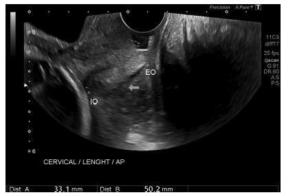
Cohort of low-risk pregnantwomen undergoing transvaginal ultrasound exams every 4 weeks, comprisingmeasurements of the cervical length and volume, the transverse and anteroposterior diameters of the cervix, and distance fromthe entrance of the uterine artery into the cervix until the internal os. The inter- and intraobserver variabilities were assessed with the linear correlation coefficient and the Student t-test. Within each period of GA, 2.5, 10, 50, 90 and 97.5 percentiles were estimated, and the variation by GA was assessed with analysis of variance for dependent samples. Mean values and Student t-test were used to compare the values stratified by control variables.
After confirming the high reproducibility of the method, 172 women followed in this cohort presented a reduction in cervical length, with an increase in volume and in the anteroposterior and transverse diameters during pregnancy. Smaller cervical lengths were associated with younger age, lower parity, and absence of previous cesarean section (C-section).
In the studied population, we observed cervical length shortening throughout pregnancy, suggesting a physiological reduction mainly in the vaginal portion of the cervix. In order to better predict pretermbirth, cervical insufficiency and premature rupture of membranes, reference curves and specific cut-off values need to be validated.

Summary
Revista Brasileira de Ginecologia e Obstetrícia. 2017;39(9):453-463
To assess the knowledge and compliance of health professionals regarding the diagnostic and treatment practices for syphilis in patients admitted for childbirth in public maternity hospitals in the city of Teresina, in the state of Piauí, Northeastern Brazil.
A cross-sectional study was performed in 2015 with obstetricians and nurses working in the public maternity hospitals in Teresina (n = 159) using a selfadministered questionnaire, with 5% of losses and 10% of refusals. The study used 21 evaluation criteria: 13 of them were related to knowledge (5 on serological tests and 8 on treatment adequacy); 8 were related to practices (3 on diagnosis, 4 on treatment, and 1 on post-test counseling). The knowledge of and compliance to the practices was estimated as the proportion of health professionals’ answers that were in agreement with Brazilian Ministry of Health protocols.
The obstetricians were in agreement with twocriteria concerning the knowledge of serological tests, one for diagnostic practices, and one for treatment practice. Among nurses, no single match between actual procedures and guidelines was observed.
Low compliance with the protocols results in missed opportunities for the diagnosis and treatment of pregnant and postpartum women and their partners. Strategies for training and integrating the various professional groups, improved data recording on prenatal cards, and greater accountability of the hospital team in managing the women’s partners are needed to overcome the barriers identified in the study and to interrupt the syphilis transmission chain.
Summary
Revista Brasileira de Ginecologia e Obstetrícia. 2017;39(8):415-423
To assess themedical doctors andmedical students’ opinion regarding the evidence and ethical background of the performance of vulvovaginal aesthetic procedures (VVAPs).
Cross-sectional online survey among 664 Portuguese medical doctors and students.
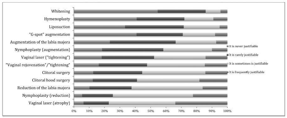
Most participants considered that there is never or there rarely is amedical reason to perform: vulvar whitening (85.9% [502/584]); hymenoplasty (72.0% [437/607]); mons pubis liposuction (71.6% [426/595]); “G-spot” augmentation (71.0% [409/576]); labia majora augmentation (66.3% [390/588]); labia minora augmentation (58.3% [326/559]); or laser vaginal tightening (52.3%[313/599]).Gynecologists and specialistsweremore likely to consider that there are no medical reasons to performVVAPs; the opposite was true for plastic surgeons and students/residents. Hymenoplasty raised ethical doubts in 51.1% (283/554) of the participants. Plastic surgeons and students/residents were less likely to raise ethical objections, while the opposite was true for gynecologists and specialists. Most considered that VVAPs could contribute to an improvement in self-esteem(92.3% [613/664]); sexual function (78.5% [521/664]); vaginal atrophy (69.9% [464/664]); quality of life (66.3% [440/664]); and sexual pain (61.4% [408/664]).
While medical doctors and students acknowledge the lack of evidence and scientific support for the performance of VVAPs, most do not raise ethical objections about them, especially if they are students or plastic surgeons, or if they have had or have considered having plastic surgery.

Summary
Revista Brasileira de Ginecologia e Obstetrícia. 2017;39(8):408-414
the aim of this study was to evaluate the pattern of human papillomavirus (HPV) detection in an 11.3-year post-vaccination period in a cohort of adolescent and young women vaccinated or not against HPV 16/18.
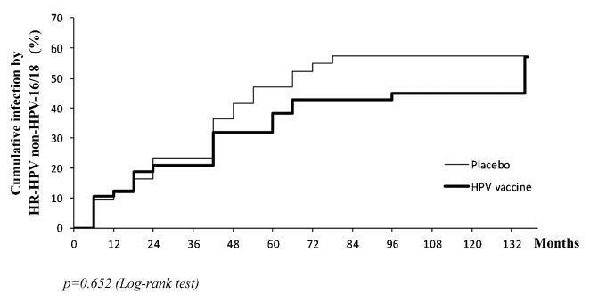
a subset of 91 women from a single center participating in a randomized clinical trial (2001-2010, NCT00689741/00120848/00518336) with HPV 16/18 AS04- adjuvanted vaccine was evaluated. All women received three doses of the HPV vaccine (n = 48) or a placebo (n = 43), and cervical samples were collected at 6-month intervals. Only in this center, one additional evaluation was performed in 2012. Up to 1,492 cervical samples were tested for HPV-DNA and genotyped with polymerase chain reaction (PCR). The vaccine group characteristics were compared by Chi-square or Fisher exact or Mann-Whitney test. The high-risk (HR)-HPV 6-month-persistent infection rate was calculated. The cumulative infection by HPV group was evaluated by the Kaplan-Meier method and the log-rank test.
the cumulative infection with any type of HPV in an 11.3-year period was 67% in the HPV vaccine group and 72% in the placebo group (p = 0.408). The longitudinal analysis showed an increase of 4% per year at risk for detection of HR-HPV (non-HPV 16/ 18) over time (p = 0.015), unrelated to vaccination. The cumulative infection with HPV 16/18 was 4% for the HPV vaccine group and 29% for the placebo group (p = 0.003). There were 43 episodes of HR-HPV 6-month persistent infection, unrelated to vaccination.
this study showed themaintenance of viral detection rate accumulating HR-HPV (non-HPV-16-18) positive tests during a long period post-vaccination, regardless of prior vaccination. This signalizes that the high number of HPV-positive testsmay be maintained after vaccination.

Summary
Revista Brasileira de Ginecologia e Obstetrícia. 2017;39(8):403-407
To determine the clinical and epidemiological characteristics of abdominal wall endometriosis (AWE), as well as the rate and recurrence factors for the disease.
A retrospective study of 52 women with AWE was performed at Universidade Estadual de Campinas from 2004 to 2014. Of the 231 surgeries performed for the diagnosis of endometriosis, 52 women were found to have abdominal wall endometriosis (AWE). The frequencies, means and standard deviations of the clinical characteristics of these women were calculated, as well as the recurrence rate of AWE. To determine the risk factors for disease recurrence, Fisher’s exact test was used.
The mean age of the patients was 30.71 ± 5.91 years. The main clinical manifestations were pain (98%) and sensation of a mass (36.5%).We observed that 94% of these women had undergone at least 1 cesarean section, and 73% had used medication for the postoperative control of endometriosis. The lesion was most commonly located in the cesarean section scar (65%). The recurrence rate of the disease was of 26.9%. All 14 women who had relapsed had surgical margins compromised in the previous surgery. There was no correlation between recurrent AWE and a previous cesarean section (p = 0.18), previous laparotomy (p = 0.11), previous laparoscopy (p = 0.12) and postoperative hormone therapy (p = 0.51).
Women with previous cesarean sections with local pain or lumps should be investigated for AWE. The recurrence of AWE is high, especially when the first surgery is not appropriate and leaves compromised surgical margins.
Summary
Revista Brasileira de Ginecologia e Obstetrícia. 2017;39(8):397-402
To describe the reproductive variables associated with different sickle cell disease (SCD) genotypes and the influence of contraceptive methods on acute painful episodes among the women with the homozygous hemoglobin S (HbSS) genotype.
A cross-sectional study was conducted between September of 2015 and April of 2016 on 158 women afflicted with SCD admitted to a hematology center in the Northeast of Brazil. The reproduction-associated variables of different SCD genotypes were assessed using the analysis of variance (ANOVA) test to compare means, and the Kruskal-Wallis test to compare medians. The association between the contraceptive method and the acute painful episodes was evaluated by the Chi-square test.
Themean age of women with SCD was 28.3 years and 86.6% were mixed or of African-American ethnicity. With respect to the genotypes, 134 women (84.8%) had HbSS genotype, 12 women (7.6%) had hemoglobin SC (HbSC) disease genotype, and 12 (7.6%) were identified with hemoglobinopathy S-beta (S-β) thalassemia. The mean age of HbSS diagnosis was lower than that of HbSC disease, the less severe formof SCD (p < 0.001). The mean age ofmenarche was 14.8 ± 1.8 years for HbSS and 12.7 ± 1.5 years for HbSC (p < 0.001). Among women with HbSS who used progestin-only contraception, 16.6% had more than 4 acute painful episodes per year. There was no statistically significant difference when compared with other contraceptive methods.
With respect to reproduction-associated variables, only the age of the menarche showed delay in HbSS when compared with HbSC. The contraceptive method used was not associated with the frequency of acute painful episodes among the HbSS women.