Summary
Revista Brasileira de Ginecologia e Obstetrícia. 2008;30(5):257-260
DOI 10.1590/S0100-72032008000500008
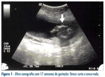
Camptomelic dysplasia belongs to a heterogeneous and rare group of lethal skeletal dysplasias, characterized by abnormal development of bones and cartilages. It is caused by a mutation in gene Sox9 (SRY-like HMG [high-mobility group] BOX 9) of chromosome 17 and it is transmitted as an autosomal dominant trait. Its main characteristics are the shortening and bowing of the long bones, principally the lower limbs. It is also associated with other severe skeletal and extraskeletal malformations. Karyotype study may reveal sex reversal. The majority of carriers die during the fetal and early neonatal periods. Ultrasound is essential to elucidate a prenatal diagnosis.

Summary
Revista Brasileira de Ginecologia e Obstetrícia. 2006;28(10):575-580
DOI 10.1590/S0100-72032006001000002
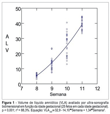
PURPOSE: To determine the values of amniotic fluid in normal fetuses during the first trimester of pregnancy by three- and bi-dimensional ultrasonography. METHODS: In a prospective longitudinal study, 25 normal fetuses were evaluated from the 8th to the 11th week of gestation. Amniotic fluid volume was measured by endovaginal ultrasonography with the three- and two-dimensional modes. The two-dimensional study consisted of volumetric determination by mathematical calculation based on an ellipsoidal shape (constant 0.52) to obtain the amniotic sac and embryo volumes. In the three-dimensional study, the amniotic fluid volume was determined by the VOCAL technique using 6, 9, 15, and 30 degrees of rotation. The amniotic fluid volume obtained by 6-degree rotations was considered to be the final result. In both modes, amniotic fluid volume was obtained by subtracting the volume of the embryo from the volume of the amniotic sac. Data were analyzed statistically for variance (ANOVA), correlation and regression analysis. The level of significance was set at p < 0.05. RESULTS: The amniotic fluid volume as measured by two-dimensional ultrasonography increased from 5.45 to 39.52 cm³ in the range from the 8th to the 11th week (ANOVA - p < 0.05). There was a correlation between gestational age and amniotic fluid volume (p < 0.001, r² = 88.3%). In the three-dimensional study, the amniotic fluid volume increased from 5.7 to 42.9 cm³ in the range from the 8th to the 11th week (ANOVA - p < 0.05), and again a correlation between gestational age and amniotic fluid volume (p < 0.001, r² = 98.1%) was observed. CONCLUSION: an increase in amniotic fluid volume occurs during the first trimester of pregnancy, as determined by the two- and three-dimensional modes. In addition, we have demonstrated that the higher the gestational age, the larger the amniotic fluid volume.

Summary
Revista Brasileira de Ginecologia e Obstetrícia. 2006;28(3):165-170
DOI 10.1590/S0100-72032006000300005
PURPOSE: to describe perinatal and obstetric characteristics of pregnant women with ultrasonographic early placental aging. METHODS: using a retrospective, descriptive, series of cases, with group comparison, the authors analyzed the data of 146 pregnant women, whose diagnosis of placental early aging (presence of grade II placenta before 32 gestational weeks or grade III, before 35 gestational weeks), and maternal-fetal conditions had been recorded in the medical charts at the "Maternidade Prof. Monteiro de Moraes", Recife, Pernambuco Brazil, from January 2000 to December 2002, where they had been attended as inpatients. The exclusion criteria were diagnoses of: premature amniorrhexis, multiple pregnancies, acute premature detachment of a normally located placenta, and fetal malformation. The clinical and obstetric complications were: hypertensive diseases, intrauterine growth restriction, changes of amniotic fluid volume, infections, maternal diabetes, falciform anemia, HIV seropositivity, drug addiction, renal lithiasis, epilepsy and bronchial asthma. In the medical records, 106 pregnant women were identified as having clinical and obstetric complications (Gwith group) and 40 as not having any of these complications (Gwithout group). For group comparisons, chi2 and exact Fisher statistical tests were used, with significance level of 0.05. RESULTS: Gwith group was associated with higher incidence of oligoamnion (27.3%), intrauterine growth restriction (44.3%) and caesarean section prior to labor (36.8%). Compared to Gwithout, the Gwith group was characterized by high incidence of: fetal death, prematurity (58.8% versus 40%), lower 5th minute Apgar index, birth weight less than 2.500g (67.9% versus 40%); small body size for gestational age (39.2% versus 10%) and more severe intercurrents events. CONCLUSIONS: perinatal prognosis does not depend upon placental early aging, but on clinical and obstetric maternal complications.
Summary
Revista Brasileira de Ginecologia e Obstetrícia. 2005;27(6):310-315
DOI 10.1590/S0100-72032005000600004
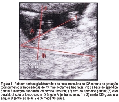
PURPOSE: to evaluate the accuracy of fetal gender prediction at 11 to 13 weeks and 6 days by measuring the anterior and posterior genital tubercle angles. MESTHODS: the anterior and posterior genital tubercle angles were measured in a midsagittal plane in 455 fetuses from 11 to 13 weeks and 6 days. The probability of a correct fetal sex prediction (confirmed after birth) was categorized in accordance with the angle measurements, gestational age and crump-rump length. The optimal accuracy cutoffs were derived from a ROC-plot. The interobserver variability was evaluated by a Bland-Altman plot. RESULTS: the correct fetal sex prediction rate increased with gestational age and crump-rump length. Using a 42-degree anterior angle as a cutoff, a correct fetal sex prediction occurred in 72% of the fetuses from 11 to 11 weeks and 6 days, 86% from 12 to 12 weeks and 6 days and 88% from 13 to 13 weeks and 6 days. Using a 24-degree posterior angle as a cutoff, a correct fetal gender prediction occurred in 70, 87 and 87%, respectively. The interobserver variability evaluation revealed a mean difference between paired measurements of 15.7 and 9 degrees for the posterior and anterior angles, respectively. CONCLUSION: the measurement of the genital tubercle angles showed a high accuracy in correctly predicting the fetal sex from the 12th week of gestation on. However, accuracy was still not high enough for clinical use in pregnancies at risk of serious X-linked diseases.
