Summary
Revista Brasileira de Ginecologia e Obstetrícia. 2005;27(6):353-356
DOI 10.1590/S0100-72032005000600010
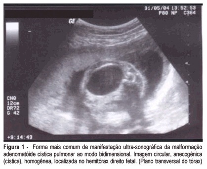
Fetal cystic adenomatoid malformation is a pulmonary developmental anomaly arising from an overgrowth of the terminal respiratory bronchioles. This is such a rare malformation, that is not always thought of as a diagnostic possibility. We present a case of pulmonary cystic adenomatoid malformation and emphasize the importance of early diagnosis and therapeutic possibilities. We also present its evolution after prenatal placement of a catheter for continuous drainage.

Summary
Revista Brasileira de Ginecologia e Obstetrícia. 2005;27(6):310-315
DOI 10.1590/S0100-72032005000600004
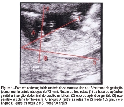
PURPOSE: to evaluate the accuracy of fetal gender prediction at 11 to 13 weeks and 6 days by measuring the anterior and posterior genital tubercle angles. MESTHODS: the anterior and posterior genital tubercle angles were measured in a midsagittal plane in 455 fetuses from 11 to 13 weeks and 6 days. The probability of a correct fetal sex prediction (confirmed after birth) was categorized in accordance with the angle measurements, gestational age and crump-rump length. The optimal accuracy cutoffs were derived from a ROC-plot. The interobserver variability was evaluated by a Bland-Altman plot. RESULTS: the correct fetal sex prediction rate increased with gestational age and crump-rump length. Using a 42-degree anterior angle as a cutoff, a correct fetal sex prediction occurred in 72% of the fetuses from 11 to 11 weeks and 6 days, 86% from 12 to 12 weeks and 6 days and 88% from 13 to 13 weeks and 6 days. Using a 24-degree posterior angle as a cutoff, a correct fetal gender prediction occurred in 70, 87 and 87%, respectively. The interobserver variability evaluation revealed a mean difference between paired measurements of 15.7 and 9 degrees for the posterior and anterior angles, respectively. CONCLUSION: the measurement of the genital tubercle angles showed a high accuracy in correctly predicting the fetal sex from the 12th week of gestation on. However, accuracy was still not high enough for clinical use in pregnancies at risk of serious X-linked diseases.

Summary
Revista Brasileira de Ginecologia e Obstetrícia. 2000;22(4):191-199
DOI 10.1590/S0100-72032000000400002
Purpose: to determine the frequency of prenatal diagnosis in newborns with gastroschisis operated at the Instituto Materno-Infantil de Pernambuco (IMIP) and to analyze its repercussions on neonatal prognosis. Patients and Methods: a cross-sectional study was carried out, including 31 cases of gastroschisis submitted to surgical correction in our service from 1995 to 1999. Prevalence risk (PR) of neonatal death and its 95% confidence interval were calculated for the presence of prenatal diagnosis and other perinatal and surgical variables. Multiple logistic regression analysis was carried out to determine the adjusted risk of neonatal death. Results: only 10 of 31 cases of gastroschisis (32.3%) had prenatal diagnosis and all were delivered at IMIP. No newborn with prenatal diagnosis was preterm but 43% of those without prenatal diagnosis were premature (p < 0,05). Birth-to-surgery interval was significantly greater in the absence of prenatal diagnosis (7.7 versus 3.8 hours). The type of surgery, need of mechanical ventilation and frequency of postoperative infection were not different between the groups. Neonatal death was more frequent in the group without prenatal diagnosis (67%) than in the group with prenatal diagnosis (20%). The main factors associated with increased risk of neonatal death were gestational age <37 weeks, absence of prenatal diagnosis, delivery in other hospitals, birth-to-surgery interval > 4 hours, staged silo surgery, need of mechanical ventilation and postoperative infection. Conclusions: prenatal diagnosis was infrequent among infants with gastroschisis and neonatal death was extremely high in its absence. It is necessary to achieve greater rates of prenatal diagnosis and to improve perinatal care in order to reduce this increased mortality.
Summary
Revista Brasileira de Ginecologia e Obstetrícia. 2004;26(5):377-382
DOI 10.1590/S0100-72032004000500006
OBJECTIVE: to describe ultrasonographic alterations in fetuses infected with Toxoplasma gondii, correlating them with neonatal prognosis. METHODS: between June 1997 and May 2003, 150 pregnant women with suspected toxiplasmosis were examined. Acute infection was confirmed in 72 (48%) of these pregnant women and congenital toxoplasmosis was diagnosed in 12 (16%) fetuses. Prenatal diagnosis was established by polymerase chain reaction in the amniotic fluid. All the patients received antiparasitic therapy. Ultrasound examination was performed every fortnight and all the infants were evaluated during their first year of life. RESULTS: ultrasonographic changes were observed in eight fetuses. All of them showed symmetric bilateral ventricular enlargement that was associated with periventricular calcifications in five cases. Other changes as hepatic calcification, hepatomegaly, polyhydramnium, and pericardial effusion were less frequent. Among these fetuses, four were stillborn and three showed sequelae (chorioretinitis and neuro-psychomotor retardation). The four fetuses that showed normal ultrasonography had a satisfactory development. CONCLUSION: There was a high incidence of ultrasonographic changes in fetuses with congenital toxoplasmosis, mainly brain damage. Other changes as hepatomegaly and pericardial effusion were less frequent and were related to a systemic infection. The prognosis of these fetuses seems to be correlated with the presence of these lesions mainly because they had high mortality ratio and among the survivors the incidence of sequelae was high. The non-symptomatic fetuses evolved in a favorable way without developing sequelae. These results highlight the value of ultrasonographic examination of these fetuses in order to establish a prognosis and allow the elaboration of a suitable post-natal procedure.
Summary
Revista Brasileira de Ginecologia e Obstetrícia. 2004;26(5):383-389
DOI 10.1590/S0100-72032004000500007
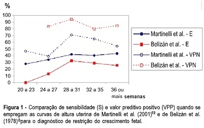
OBJECTIVE: to evaluate the measurement of uterine height in order to predict fetal growth restriction (FGR), according to a local curve. METHODS: from July 2000 to February 2003, 238 high-risk pregnant women were submitted to uterine height measurements between the 20th and the 42nd week of gestation. The gestational age of all the women was well known, confirmed by early ultrasound. Fifty (21%) women gave birth to infants considered small for their gestational age. The measures were performed by a single observer, who took 1617 uterine height measurements, from the upper border of the symphysis pubis to the fundus uteri, using tape measurement. The diagnosis of FGR was confirmed after birth according to the Ramos's curve. The women were divided into two groups according to their infant's birth weight and the data were statistically analyzed by the Fisher's exact test or Kruskal-Wallis's test. The sensitivity (SE), specificity (SP), positive predictive value (PPV), and negative predictive value (NPV) were calculated. The test for two proportions with normal approximation was performed to analyze the continuous variables. RESULTS: one measurement below the 10th percentile, according to gestational age, resulted in SE = 78.0%, SP = 77.1%, PPV = 47.6%, and NPV = 88.8% for the identification of FGR. If one measurement was below the 5th percentile, the SE, SP, PPV, and NPV were 64.0, 89.9, 62.7 and 90.4%, respectively. CONCLUSIONS: one measurement below the 10th percentile for the gestational age, according to the local curve, proved to be a good predictor of FGR.

Summary
Revista Brasileira de Ginecologia e Obstetrícia. 2000;22(6):365-371
DOI 10.1590/S0100-72032000000600007
Purpose: to evaluate the accuracy of prenatal ultrasound in the diagnosis of nephrouropathies. Methods: the authors followed-up 127 pregnancies referred to the Fetal Medicine Center of UFMG with suspicion of these anomalies. Fetal biometry, growth, vitality, and associated malformations were evaluated. Finally, a detailed description of the renal system was made to define the prenatal morphologic diagnosis of the malformations to be compared with the postnatal diagnosis. Results: based on the kappa index (statistical method that measures the concordance between different measurements, methods or measurement instruments: below 0.40, poor agreement; between 0.40 and 0.75, good agreement; above 0.75, excellent ageement), the authors found an excellent concordance (kappa index 0.95). Among the 127 cases, there were only 9 misdiagnoses, all of them of obstructive uropathies: 6 cases showed different obstruction levels after delivery and in three cases there were confounding diagnosis with multicystic kidney. Conclusions: the detailed ultrasonographic description of the renal system is a good method for prenatal diagnosis of the fetal nephropathies, allowing some options to modify the outcome of these fetuses, like to send them to specialized centers, to anticipate delivery and even to apply intrauterine therapy, in order to preserve the renal function. Serial echography and amnioinfusion can be used to improve the precision of prenatal diagnosis.
Summary
Revista Brasileira de Ginecologia e Obstetrícia. 2000;22(7):421-428
DOI 10.1590/S0100-72032000000700004
Purpose: to evaluate 24 cases of gastroschisis, in relation to the prognostic factors that interfered with postnatal outcome. Patients and Method: twenty-four pregnancies with fetal prenatal ultrasound diagnosis of gastroschisis, during an 8-year period, were analyzed. Gastroschisis was classified into isolated, when there were no other structural abnormalities, or associated, when other abnormalities were present. For both groups the following parameters were examined: ultrasound bowel dilatation (>18 mm), obstetric complications and postnatal outcome. Nonparametric Mann-Whitney and exact Fisher's tests were used for statistical analyses. Results: in 9 cases (37.5%) gastroschisis was associated with other abnormalities, and in 15 cases it was isolated (62.5%). All cases of associated gastroschisis had a letal prognosis, therefore the overall mortality rate was 60.8%. In the group of isolated gastroschisis, all were born alive and were submitted to surgery, but the survival rate after surgical correction was 60%. The median gestational age at birth was 35 weeks and birth weight 2,365 grams. Premature delivery was observed in 10 cases, mainly as a consequence of obstetric complication. Two newborns were small for gestational age, and only 3 had birth weight >2,500 grams. Oligohydramnios was found in 46.6% and it was more frequent in the group of postnatal death (66.7%). Ultrasound assessment of bowel showed bowel dilatation in 86.6%, however, without relation to the prognosis and postnatal bowel findings. There was no significant difference between gestational age at birth and birth weight comparing the survivor and postnatal death groups. Conclusions: isolated gastroschisis had a better prognosis when compared to associated, therefore this prenatal differentiation is important. Isolated gastroschisis was often associated with prematurity, small birth weight and obstetric complications. Prenatal diagnosis allows better monitoring of fetal and obstetric conditions. Delivery should be at term, unless presenting with obstetric complications.
Summary
Revista Brasileira de Ginecologia e Obstetrícia. 2001;23(1):31-37
DOI 10.1590/S0100-72032001000100005
Purpose: to evaluate the prognosis of fetal omphalocele after prenatal diagnosis. Methods: fifty-one cases with prenatal diagnosis of fetal omphalocele were divided into three groups: group 1, isolated omphalocele; group 2, omphalocele associated with structural abnormalities and normal karyotype; group 3, omphalocele with abnormal karyotype. The data were analyzed for overall survival rate and postsurgery survival, considering associated malformations, gestational age at delivery, birth weight and size of omphalocele. Results: group 1 corresponded to 21% (n = 11), group 2, 55% (n = 28) and group 3,24% (n = 12). All of Group 3 died, and trisomy 18 was the most frequent chromosomal abnormality. The survival rate was 80% for group 1 and 25% for group 2. Sixteen cases underwent surgery (10 isolated and 6 associated), 81% survived (8 isolated and 5 associated). The median birth weight was 3,140 g and 2,000 g for survivals and non-survivals after surgery, respectively (p = 0.148), and the corresponding gestational age at delivery was 37 and 36 weeks (p = 0.836). The ratio of omphalocele/abdominal circumference decreased with gestation, 0.88 between 25-29 weeks and 0.65 between 30-35 weeks (p = 0.043). The size of omphalocele was not significantly different between the 3 groups (p = 0.988), and it was not associated to postsurgery prognosis (p = 0.553). Conclusion: the overall and postsurgery survival rates were 25 and 81%, respectively. Associated malformations were the main prognostic factor in prenatally diagnosed omphaloceles, since they are associated with prematurity and low birth weight.