Summary
Revista Brasileira de Ginecologia e Obstetrícia. 2014;36(3):139-141
DOI 10.1590/S0100-72032014000300008
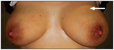
Mondor's disease is a rare entity characterized by sclerosing thrombophlebitis classically involving one or more of the subcutaneous veins of the breast and anterior chest wall. It is usually a self-limited, benign condition, despite of rare cases of association to cancer. We present the case of a 32 year-old female, breast-feeding, who went to emergency due to left mastalgia for the past week. She was taking antibiotics and non-steroidal anti-inflammatory drugs, previously prescribed for suspicious of mastitis, for three days, with no clinical improvement. Physical examination showed an enlarged left breast, an axillary lump and a painful cord-like structure in the upper outer quadrant of the same breast. Ultrasound scan showed a markedly dilated superficial vein in the upper outer quadrant of left breast. The patient was given a ventropic therapy and was kept in anti-inflammatory, with progressive pain improvement. Ultrasound control was performed after four weeks, showing reperfusion.

Summary
Revista Brasileira de Ginecologia e Obstetrícia. 2013;35(8):368-372
DOI 10.1590/S0100-72032013000800006
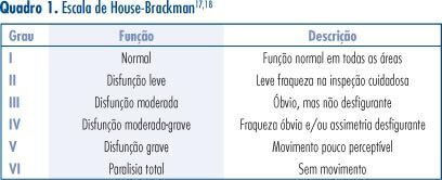
PURPOSE: To compare the degree of peripheral facial palsy of pregnant women and puerperae at admission and at discharge and to evaluate related factors. METHODS: Retrospective, cross-sectional study, with analysis of medical records of pregnant and postpartum women with facial palsy, over a period of 12 months, with application of a standardized protocol for patient evaluation and of the House-Brackmann scale on the occasion of the first visit and at discharge. RESULTS: Six patients were identified, mean age of 22.6 years. Five cases were classified as stage IV and one as stage II on the House-Brackmann scale, being two of them puerperae and four pregnant. All showed improvement on the House-Brackmann scale. CONCLUSION: The Bell's palsy has a good prognosis even in pregnant and postpartum women, being important to perform the correct treatment to reduce the sequelae in this group identified as more susceptible to peripheral facial palsy.

Summary
Revista Brasileira de Ginecologia e Obstetrícia. 2013;35(1):10-15
DOI 10.1590/S0100-72032013000100003
PURPOSES: To investigate the effect of an individualized and supervised exercise program for the pelvic floor muscles (PFM) in the postpartum period of multiparous women, and to verify the correlation between two methods used to assess PFM strength. METHODS: An open clinical trial was performed with puerperal, multiparous women aged 18 to 35 years. The sample consisted of 23 puerperal women divided into two groups: Intervention Group (IG, n=11) and Control Group (CG, n=12). The puerperal women in IG participated in an eight-week PFM exercise program, twice a week. The puerperal women in CG did not receive any recommendations regarding exercise. PFM strength was assessed using digital vaginal palpation and a perineometer. The statistical analysis was performed using the following tests: Fisher's exact, c², Student's t, Kolmogorov-Smirnov for two samples, and Pearson's correlation coefficient. Significance was defined as p<0.05. RESULTS: The participants' mean age was 24±4.5 years in IG and 25.3±4 years in CG (p=0.4). After the exercise program, a significant difference was found between the groups in both modalities of muscle strength assessment (p<0.001). The two muscle strength assessment methods showed a significant correlation in both assessments (1st assessment: r=0.889, p<0.001; 2nd assessment: r=0.925, p<0.001). CONCLUSIONS: The exercise program promoted a significant improvement in PFM strength. Good correlation was observed between digital vaginal palpation and a perineometer, which indicates that vaginal palpation can be used in clinical practice, since it is an inexpensive method that demonstrated significant correlation with an objective method, i.e. the use of a perioneometer.
Summary
Revista Brasileira de Ginecologia e Obstetrícia. 2012;34(4):158-163
DOI 10.1590/S0100-72032012000400004
PURPOSE: To verify cervical length using transvaginal ultrasonography in pregnant women between 28 and 34 weeks of gestation, correlating it with the latent period and the risk of maternal and neonatal infections. METHODS: 39 pregnant women were evaluated and divided into groups based on their cervical length, using 15, 20 and 25 mm as cut-off points. The latency periods evaluated were three and seven days. Included were pregnant women with live fetuses and gestational age between 28 and 34 weeks, with a confirmed diagnosis on admission of premature rupture of membranes. Patients with chorioamnionitis, multiple gestation, fetal abnormalities, uterine malformations (bicornus septate and didelphic uterus), history of previous surgery on the cervix (conization and cerclage) and cervical dilation greater than 2 cm in nulliparous women and 3 cm in multiparae were excluded from the study. RESULTS: A <15 mm cervical length was found to be highly related to a latency period of up to 72 hours (p=0.008). A <20 mm cervical length was also associated with a less than 72 hour latency period (p=0.04). A <25 mm cervical length was not found to be statistically associated with a 72 hour latency period (p=0,12). There was also no significant correlation between cervical length and latency period and maternal and neonatal infection. CONCLUSION: The presence of a short cervix (<15 mm) was found to be related to a latency period of less than 72 hours, but not to maternal or neonatal infections.
Summary
Revista Brasileira de Ginecologia e Obstetrícia. 2011;33(9):252-257
DOI 10.1590/S0100-72032011000900006
PURPOSE: To describe and compare the phases of stress of primiparae in the third trimester of pregnancy and postpartum, associating them with the occurrence of postpartum depression. METHODS: The study consisted of two stages (Stage 1 and Stage 2), characterized as longitudinal research. Ninety-eight primiparae participated in Stage 1, and 64 of them participated in Stage 2. In Stage 1, data were collected in the third trimester of pregnancy, and in Stage 2, at least 45 days after delivery. The Stress Symptoms Inventory Lipp (ISSL) was applied in Stage 1 and an interview was held to characterize the sample. In Stage 2, we applied again the ISSL and also the EPDS (Edinburgh Postnatal Depression Scale). Data were analyzed using SPSS for Windows®, version 17.0. The statistical analyses were performed using the Student’s t-test and the Spearman p. RESULTS: Seventy-eight percent of the participants showed significant signs of stress in the third quarter and 63% of them during the postpartum period, with a significant difference in the stress occurring in the third trimester and postpartum (t=2.20, p=0.03). There was also a correlation between the stress occurring during pregnancy and in the puerperium and the manifestation of postpartum depression (p<0.001). CONCLUSION: More than half of the women experience significant stress signs during both pregnancy and the postpartum period. However, the frequency of onset of significant symptoms of stress was higher during pregnancy than during the puerperium. These results seem to be closely related to the manifestation of postpartum depression, indicating the relationship between stress and postpartum depression.
Summary
Revista Brasileira de Ginecologia e Obstetrícia. 2011;33(4):188-195
DOI 10.1590/S0100-72032011000400007
PURPOSE: to evaluate the prevalence of urinary symptoms and association between pelvic floor muscle function and urinary symptoms in primiparous women 60 days after vaginal delivery with episiotomy and cesarean section after labor. METHODS: a cross-sectional analysis was conducted on women from an out patient clinic in São Paulo state, Brazil, 60 days after delivery. Pelvic floor muscle function was assessed by surface electromyography (basal tone, maximal voluntary contraction and mean sustained contraction) and by a manual muscle test (grades 0-5). In an interview, the urinary symptoms were identified and women with difficulty to understand, with motor/neurological impairment, pelvic surgery, diabetes, restriction for vaginal palpation and practicing exercises forpelvic floor muscles were excluded. The χ2 and Fisher Exact test were used to compare proportions and the Mann-Whitney test was used to analyze mean differences. RESULTS: 46 primiparous were assessed on average 63.7 days postpartum. The most prevalent symptoms were nocturia (19.6%), urgency (13%) and increased daytime urinary frequency (8.7%). Obese and overweight women had 4.6 times more of these symptoms (PR=4.6 [95%CI; 1.2-18.6; p value=0.0194]). Stress urinary incontinence was the most prevalent incontinence (6.5%). The mean values found for the basic tone, maximal voluntary contraction and sustained contraction were: 3 µV, 14.6 µV and 10.3 µV. Most of the women (56.5%) had grade 3 muscular strength. There was no association between urinary symptoms and pelvic floor muscle function. CONCLUSION: the prevalence of urinary symptoms was low 60 days postpartum and there was no association between pelvic floor muscle function and urinary symptoms.
Summary
Revista Brasileira de Ginecologia e Obstetrícia. 2010;32(7):340-345
DOI 10.1590/S0100-72032010000700006