Summary
Revista Brasileira de Ginecologia e Obstetrícia. 2014;36(5):205-210
DOI 10.1590/S0100-7203201400050004
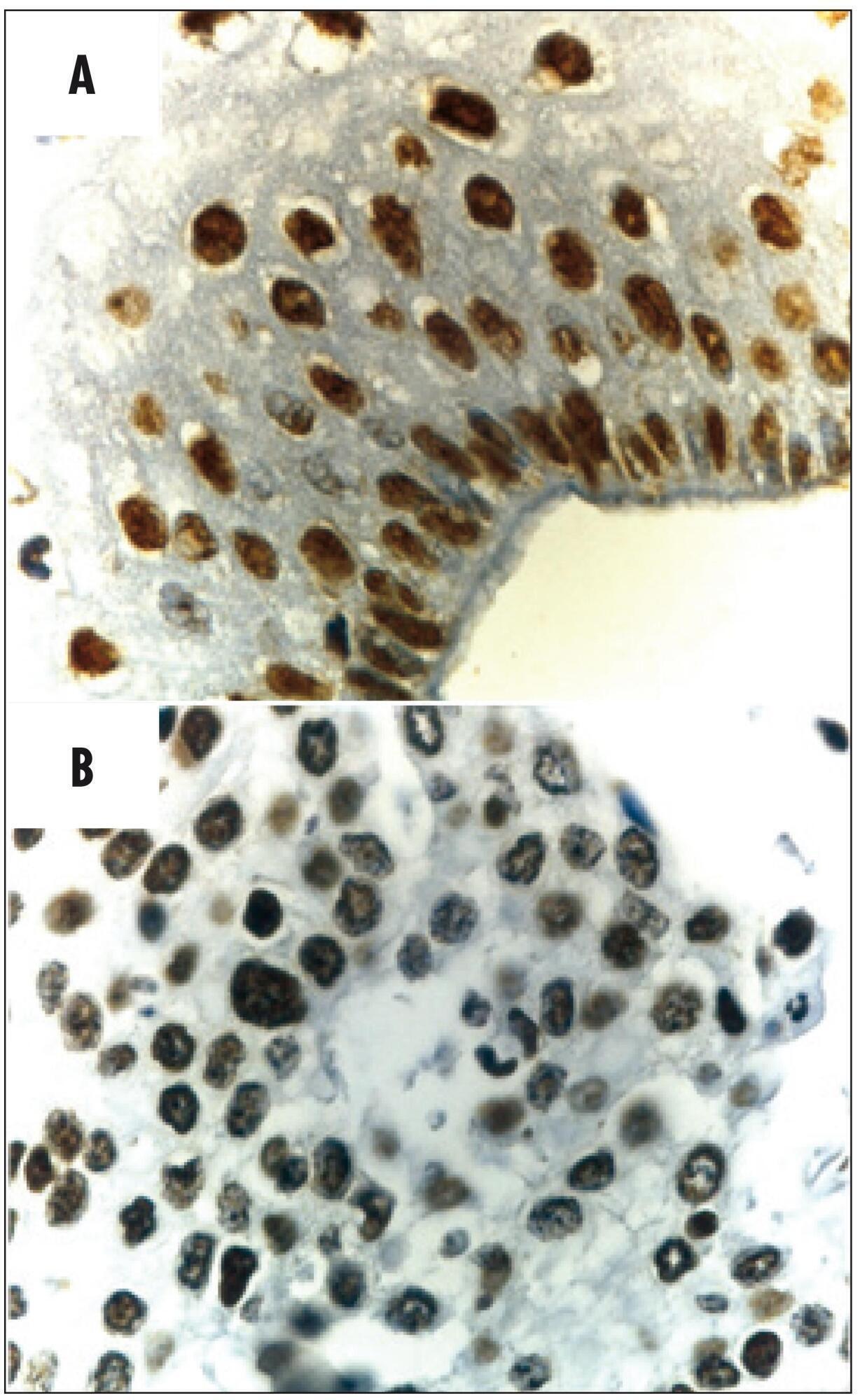
To investigate protein expression and mutations in phosphatase and tensin homolog (PTEN) in patients with stage IB cervical squamous cell carcinoma (CSCC) and the association with clinical-pathologic features, tumor p53 expression, cell proliferation and angiogenesis.
Women with stage IB CSCC (n=20 - Study Group) and uterine myoma (n=20 - Control Group), aged 49.1±1.7 years (mean±standard deviation, range 27-78 years), were prospectively evaluated. Patients with cervical cancer were submitted to Piver-Rutledge class III radical hysterectomy and pelvic lymphadenectomy and patients in the Control Group underwent vaginal hysterectomy. Tissue samples from the procedures were stained with hematoxylin and eosin for histological evaluation. Protein expression was detected by immunohistochemistry. Staining for PTEN, p53, Ki-67 and CD31 was evaluated. The intensity of PTEN immunostaining was estimated by computer-assisted image analysis, based on previously reported protocols. Data were analyzed using the Student's t-test to evaluate significant differences between the groups. Level of significance was set at p<0.05.
The PTEN expression intensity was lower in the CSCC group than in the Control (benign cervix) samples (150.5±5.2 versus 204.2±2.6; p<0.001). Our study did not identify any mutations after sequencing all nine PTEN exons. PTEN expression was not associated with tumor expression of p53 (p=0.9), CD31 (p=0.8) or Ki-67 (p=0.3) or clinical-pathologic features in patients with invasive carcinoma of the cervix.
Our findings demonstrate that the PTEN protein expression is significantly diminished in CSCC.
Summary
Revista Brasileira de Ginecologia e Obstetrícia. 2014;36(5):205-210
DOI 10.1590/S0100-7203201400050004
To investigate protein expression and mutations in phosphatase and tensin homolog (PTEN) in patients with stage IB cervical squamous cell carcinoma (CSCC) and the association with clinical-pathologic features, tumor p53 expression, cell proliferation and angiogenesis.
Women with stage IB CSCC (n=20 - Study Group) and uterine myoma (n=20 - Control Group), aged 49.1±1.7 years (mean±standard deviation, range 27-78 years), were prospectively evaluated. Patients with cervical cancer were submitted to Piver-Rutledge class III radical hysterectomy and pelvic lymphadenectomy and patients in the Control Group underwent vaginal hysterectomy. Tissue samples from the procedures were stained with hematoxylin and eosin for histological evaluation. Protein expression was detected by immunohistochemistry. Staining for PTEN, p53, Ki-67 and CD31 was evaluated. The intensity of PTEN immunostaining was estimated by computer-assisted image analysis, based on previously reported protocols. Data were analyzed using the Student's t-test to evaluate significant differences between the groups. Level of significance was set at p<0.05.
The PTEN expression intensity was lower in the CSCC group than in the Control (benign cervix) samples (150.5±5.2 versus 204.2±2.6; p<0.001). Our study did not identify any mutations after sequencing all nine PTEN exons. PTEN expression was not associated with tumor expression of p53 (p=0.9), CD31 (p=0.8) or Ki-67 (p=0.3) or clinical-pathologic features in patients with invasive carcinoma of the cervix.
Our findings demonstrate that the PTEN protein expression is significantly diminished in CSCC.
Summary
Revista Brasileira de Ginecologia e Obstetrícia. 2013;35(9):407-412
DOI 10.1590/S0100-72032013000900005
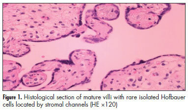
PURPOSE: In placentas from uncomplicated pregnancies, Hofbauer cells either disappear or become scanty after the fourth to fifth month of gestation. Immunohistochemistry though, reveals that a high percentage of stromal cells belong to Hofbauer cells. The aim of this study was to investigate the changes in morphology and density of Hofbauer cells in placentas from normal and pathological pregnancies. METHODS: Seventy placentas were examined: 16 specimens from normal term pregnancies, 10 from first trimester's miscarriages, 26 from cases diagnosed with chromosomal abnormality of the fetus, and placental tissue specimens complicated with intrauterine growth restriction (eight) or gestational diabetes mellitus (10). A histological study of hematoxylin-eosin (HE) sections was performed and immunohistochemical study was performed using the markers: CD 68, Lysozyme, A1 Antichymotrypsine, CK-7, vimentin, and Ki-67. RESULTS: In normal term pregnancies, HE study revealed Hofbauer cells in 37.5% of cases while immunohistochemistry revealed in 87.5% of cases. In first trimester's miscarriages and in cases with prenatal diagnosis of fetal chromosomal abnormalities, both basic and immunohistochemical study were positive for Hofbauer cells. In pregnancies complicated with intrauterine growth restriction or gestational diabetes mellitus, a positive immunoreaction was observed in 100 and 70% of cases, respectively. CONCLUSIONS: Hofbauer cells are present in placental villi during pregnancy, but with progressively reducing density. The most specific marker for their detection seems to be A1 Antichymotrypsine. It is remarkable that no mitotic activity of Hofbauer cells was noticed in our study, as the marker of cellular multiplication Ki-67 was negative in all examined specimens.
Summary
Revista Brasileira de Ginecologia e Obstetrícia. 2013;35(9):407-412
DOI 10.1590/S0100-72032013000900005
PURPOSE: In placentas from uncomplicated pregnancies, Hofbauer cells either disappear or become scanty after the fourth to fifth month of gestation. Immunohistochemistry though, reveals that a high percentage of stromal cells belong to Hofbauer cells. The aim of this study was to investigate the changes in morphology and density of Hofbauer cells in placentas from normal and pathological pregnancies. METHODS: Seventy placentas were examined: 16 specimens from normal term pregnancies, 10 from first trimester's miscarriages, 26 from cases diagnosed with chromosomal abnormality of the fetus, and placental tissue specimens complicated with intrauterine growth restriction (eight) or gestational diabetes mellitus (10). A histological study of hematoxylin-eosin (HE) sections was performed and immunohistochemical study was performed using the markers: CD 68, Lysozyme, A1 Antichymotrypsine, CK-7, vimentin, and Ki-67. RESULTS: In normal term pregnancies, HE study revealed Hofbauer cells in 37.5% of cases while immunohistochemistry revealed in 87.5% of cases. In first trimester's miscarriages and in cases with prenatal diagnosis of fetal chromosomal abnormalities, both basic and immunohistochemical study were positive for Hofbauer cells. In pregnancies complicated with intrauterine growth restriction or gestational diabetes mellitus, a positive immunoreaction was observed in 100 and 70% of cases, respectively. CONCLUSIONS: Hofbauer cells are present in placental villi during pregnancy, but with progressively reducing density. The most specific marker for their detection seems to be A1 Antichymotrypsine. It is remarkable that no mitotic activity of Hofbauer cells was noticed in our study, as the marker of cellular multiplication Ki-67 was negative in all examined specimens.
Summary
Revista Brasileira de Ginecologia e Obstetrícia. 2010;32(8):374-380
DOI 10.1590/S0100-72032010000800003
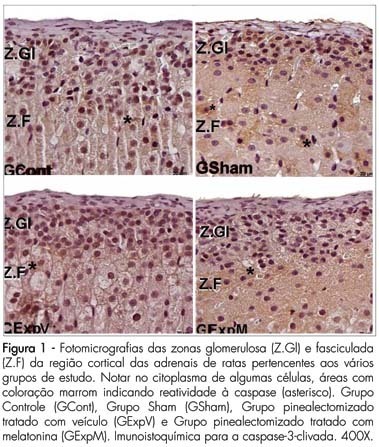
PURPOSE: to evaluate the reactivity of VEGF-A and cleaved caspase-3 in the adrenal gland cortex of female pinealectomized rats treated with melatonin. METHODS: forty adult female rats were divided into 4 groups (G) of 10 animals: GI - no surgical intervention, with vehicle administration; GII - sham pinealectomized with vehicle administration; GIII - pinealectomized with vehicle administration; GIV - pinealectomized with melatonin administration (10 µg/animal) during the night. After 60 days of treatment, all animals were anesthetized, and the adrenal glands were removed and fixed in 10% formaldehyde (phosphate buffered) for histological processing and paraffin embedding. Sections (5 µm thick) were collected on silanized slides and submitted to imunnohistochemical methods for the detection of cleaved caspase-3 (apoptosis) and of vascular endothelial growth factor (VEGF-A) in the adrenal cortex. The data obtained were submitted to analysis of variance (ANOVA) complemented by the Tukey-Kramer test (p<0.05). RESULTS: reactivity to cleaved Caspase-3 was noted in the zona glomerulosa of the adrenal glands in all studied groups. There were no significant differences in the zona glomerulosa; however, the zona fasciculata (15.51±3.12*, p<0.05) and the zona reticularis (8.11±1.90*, p<0.05) presented the smallest percentage of apoptosis in the pinealectomized group (GIII). The reactivity to the VEGF-A was stronger in the zona glomerulosa and weaker in the zona reticularis in all groups. We found a stronger VEGF-A reactivity in the zona fasciculata in the pinealectomized group (GIII). CONCLUSIONS: the pineal gland affects the arrangement of the zona glomerulosa and reticularis of the adrenal glands, which are related to the production of sex hormones.
Summary
Revista Brasileira de Ginecologia e Obstetrícia. 2010;32(8):374-380
DOI 10.1590/S0100-72032010000800003
PURPOSE: to evaluate the reactivity of VEGF-A and cleaved caspase-3 in the adrenal gland cortex of female pinealectomized rats treated with melatonin. METHODS: forty adult female rats were divided into 4 groups (G) of 10 animals: GI - no surgical intervention, with vehicle administration; GII - sham pinealectomized with vehicle administration; GIII - pinealectomized with vehicle administration; GIV - pinealectomized with melatonin administration (10 µg/animal) during the night. After 60 days of treatment, all animals were anesthetized, and the adrenal glands were removed and fixed in 10% formaldehyde (phosphate buffered) for histological processing and paraffin embedding. Sections (5 µm thick) were collected on silanized slides and submitted to imunnohistochemical methods for the detection of cleaved caspase-3 (apoptosis) and of vascular endothelial growth factor (VEGF-A) in the adrenal cortex. The data obtained were submitted to analysis of variance (ANOVA) complemented by the Tukey-Kramer test (p<0.05). RESULTS: reactivity to cleaved Caspase-3 was noted in the zona glomerulosa of the adrenal glands in all studied groups. There were no significant differences in the zona glomerulosa; however, the zona fasciculata (15.51±3.12*, p<0.05) and the zona reticularis (8.11±1.90*, p<0.05) presented the smallest percentage of apoptosis in the pinealectomized group (GIII). The reactivity to the VEGF-A was stronger in the zona glomerulosa and weaker in the zona reticularis in all groups. We found a stronger VEGF-A reactivity in the zona fasciculata in the pinealectomized group (GIII). CONCLUSIONS: the pineal gland affects the arrangement of the zona glomerulosa and reticularis of the adrenal glands, which are related to the production of sex hormones.
Summary
Revista Brasileira de Ginecologia e Obstetrícia. 2009;31(2):90-93
DOI 10.1590/S0100-72032009000200007
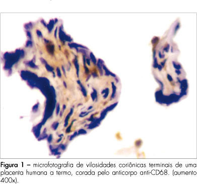
PURPOSE: to verify the amount of CD68+ cells in chorionic villosities in placentae from gestations submitted or not to labor. METHODS: transversal study with healthy near-term pregnant women, among whose placentae, 31 have been examined by immunohistochemical technique. Twenty placentae were obtained after vaginal delivery (VAGG) and eleven after elective cesarean sections (CESG). Slides were prepared with chorionic villosities samples and labeled with anti-CD68 antibody, specific for macrophages. Labeled and nonlabeled cells were counted inside the villosities. Non-parametric statistical tests were used for the analysis. RESULTS: among the 6,424 cells counted in the villosities' stroma from the 31 placentae, 1,135 cells (17.6%) were stained by the CD68+. The mean of cells labeled by the anti-CD68 was 22±18 for the VAGG group and 20±16 for the CESG, in each placentary sample. CONCLUSIONS: there were no significant differences in the percentage of macrophages (CD68+) in association with labor.
Summary
Revista Brasileira de Ginecologia e Obstetrícia. 2009;31(2):90-93
DOI 10.1590/S0100-72032009000200007
PURPOSE: to verify the amount of CD68+ cells in chorionic villosities in placentae from gestations submitted or not to labor. METHODS: transversal study with healthy near-term pregnant women, among whose placentae, 31 have been examined by immunohistochemical technique. Twenty placentae were obtained after vaginal delivery (VAGG) and eleven after elective cesarean sections (CESG). Slides were prepared with chorionic villosities samples and labeled with anti-CD68 antibody, specific for macrophages. Labeled and nonlabeled cells were counted inside the villosities. Non-parametric statistical tests were used for the analysis. RESULTS: among the 6,424 cells counted in the villosities' stroma from the 31 placentae, 1,135 cells (17.6%) were stained by the CD68+. The mean of cells labeled by the anti-CD68 was 22±18 for the VAGG group and 20±16 for the CESG, in each placentary sample. CONCLUSIONS: there were no significant differences in the percentage of macrophages (CD68+) in association with labor.
Summary
Revista Brasileira de Ginecologia e Obstetrícia. 2009;31(2):54-60
DOI 10.1590/S0100-72032009000200002
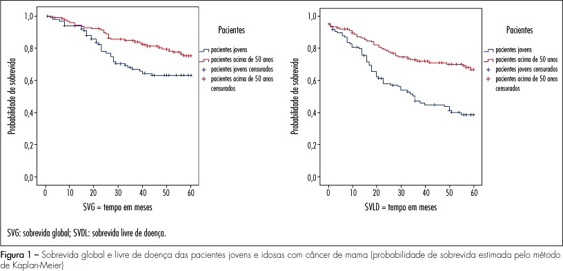
PURPOSE: the objective of this study was to evaluate the clinical, pathological and molecular characteristics in very young women and postmenopausal women with breast cancer. METHODS: we selected 106 cases of breast cancer of very young women (<35 years) and 130 cases of postmenopausal women. We evaluated clinical characteristics of patients (age at diagnosis, ethnic group, family history of breast cancer, staging, presence of distant metastases, overall and disease-free survival), pathological characteristics of tumors (tumor size, histological type and grade, axillary lymph nodes status) and expression of molecular markers (hormone receptors, HER2, p53, p63, cytokeratins 5 and 14, and EGFR), using immunohistochemistry and tissue microarray. RESULTS: when comparing clinicopathologic variables between the age groups, younger women demonstrated greater frequency of nulliparity (p=0.03), larger tumors (p<0.000), higher stage disease (p=0.01), lymph node positivity (p=0.001), and higher grade tumors (p=0.004). Most of the young patients received chemotherapy (90.8%) and radiotherapy (85.2%) and less tamoxifen therapy (31.5%) comparing with postmenopausal women. Lower estrogen receptor positivity 49.1% (p=0.01) and higher HER2 overexpression 28.7% (p=0.03) were observed in young women. In 32 young patients (29.6%) and in 20% of the posmenopausal women, the breast carcinomas were of the triple-negative phenotype (p=0.034). In 16 young women (50%) and in 10 postmenopausal women (7.7%), the tumors expressed positivity for cytokeratin 5 and/or 14, basal phenotype (p=0.064). Systemic metastases were detected in 55.3% of the young women and in 39.2% of the postmenopausal women. Breast cancer overall survival and disease-free survival in five years were, respectively, 63 and 39% for young women and 75 and 67% for postmenopausal women. CONCLUSIONS: breast cancer arising in very young women showed negative clinicobiological characteristics and more aggressive tumors.
Summary
Revista Brasileira de Ginecologia e Obstetrícia. 2009;31(2):54-60
DOI 10.1590/S0100-72032009000200002
PURPOSE: the objective of this study was to evaluate the clinical, pathological and molecular characteristics in very young women and postmenopausal women with breast cancer. METHODS: we selected 106 cases of breast cancer of very young women (<35 years) and 130 cases of postmenopausal women. We evaluated clinical characteristics of patients (age at diagnosis, ethnic group, family history of breast cancer, staging, presence of distant metastases, overall and disease-free survival), pathological characteristics of tumors (tumor size, histological type and grade, axillary lymph nodes status) and expression of molecular markers (hormone receptors, HER2, p53, p63, cytokeratins 5 and 14, and EGFR), using immunohistochemistry and tissue microarray. RESULTS: when comparing clinicopathologic variables between the age groups, younger women demonstrated greater frequency of nulliparity (p=0.03), larger tumors (p<0.000), higher stage disease (p=0.01), lymph node positivity (p=0.001), and higher grade tumors (p=0.004). Most of the young patients received chemotherapy (90.8%) and radiotherapy (85.2%) and less tamoxifen therapy (31.5%) comparing with postmenopausal women. Lower estrogen receptor positivity 49.1% (p=0.01) and higher HER2 overexpression 28.7% (p=0.03) were observed in young women. In 32 young patients (29.6%) and in 20% of the posmenopausal women, the breast carcinomas were of the triple-negative phenotype (p=0.034). In 16 young women (50%) and in 10 postmenopausal women (7.7%), the tumors expressed positivity for cytokeratin 5 and/or 14, basal phenotype (p=0.064). Systemic metastases were detected in 55.3% of the young women and in 39.2% of the postmenopausal women. Breast cancer overall survival and disease-free survival in five years were, respectively, 63 and 39% for young women and 75 and 67% for postmenopausal women. CONCLUSIONS: breast cancer arising in very young women showed negative clinicobiological characteristics and more aggressive tumors.
Summary
Revista Brasileira de Ginecologia e Obstetrícia. 2008;30(9):432-436
DOI 10.1590/S0100-72032008000900002
PURPOSE: sentinel lymph node biopsy in early-stage breast cancer patients has been substituting the total axillary lymph node is presented dissection. The technique of processing the sentinel lymph node and the aim of this study was to investigate the efficacy of occult metastasis identification based on the standard histological and immunohistochemical examination. METHODS: between 2002 and 2005, 266 sentinel lymph nodes were harvested from axillary biopsy of 170 patients with early stage breast cancer. All lymph nodes were considered to be negative according to standard intra-operative cytological assessment. Lymph nodes were transversally sectioned in four or five slices and embedded in paraffin blocks. Two paraffin-embedded tissue sections with 4 µm in thickness were mounted on glass slides and stained with hematoxylin-eosin and immunoperoxidase (cytokeratin AE1/AE3) techniques. RESULTS: standard histological examination identified metastasis in 22 patients (12.9%) and micrometastatic disease was observed in six of these patients (3.5%). The immunohistochemical examination identified metastatic disease in 16 patients (9.4%). Among them, isolated tumor cells were observed in 11 (6.5%) and micrometastases were identified in five (2.9%). CONCLUSIONS: the association of the standard histological examination and immunohistochemical technique increases the chances of sentinel lymph node metastasis identification.
Summary
Revista Brasileira de Ginecologia e Obstetrícia. 2008;30(6):287-293
DOI 10.1590/S0100-72032008000600004
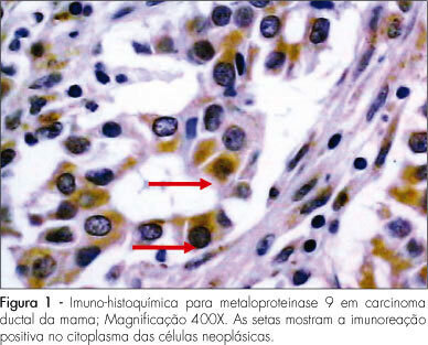
PURPOSE: to analyze the expression of matrix metalloproteinase-9 (MMP-9) and of vascular endothelial growth factor (EVGF) in a group of patients with primary breast cancer, and correlate them to one another and with other prognostic indicators. METHODS: transversal study that has analyzed the expression of MMP-9 and of VEGF in 88 consecutive cases of primary breast tumors. The samples were obtained from patients with primary breast cancer, submitted to surgical treatment in the Clinical Hospital of Porto Alegre of the Universidade Federal do Rio Grande do Sul, from January 2000 to December 2004. An immunohistochemical technique has been applied, using the avidin-biotin-peroxidase complex to evaluate the antigen immunoreactions in the tumors. The qualitative expression of proteins has been assessed through the observation of the brownish stain intensity of antibodies in the cytoplasm of malignant cells, when at least one of the tumoral cells presented clear and unequivocal staining with each of those markers. To determine the qualitative score (0=absent, 1=weak, 2=average and 3=strong), the stronger cytoplasmatic staining intensity on the glass slide has been taken into consideration, independently of the stained cells. The quantitative expression was determined by the average percentage of stained cells, observed in at least ten microscopic fields. The MMP-9 and VEGF final quantification expression has been done by the application of the HSCORE=Σ[(I+1)]xPC, where I and PC represent the staining intensity and the percentage of stained cells, respectively. RESULTS: MMP-9 and VEGF presented a significant correlation in the tumors studied. The final expression has shown a median score of 180 and 190, respectively. When MMP-9 and VEGF expression were compared with the variables "age", "tumoral diameter", "histological type", "histological grade", "axillary lymph node" and "vascular invasion", it was impossible to find any significant correlation. Compared to one another, MMP-0 and VEGF have presented a positive correlation (rho=0.23; p=0.03). The axillary lymph node positivity has presented a positive correlation with the larger tumoral diameter (2.7±1.1 cm; p<0.01) and with the presence of vascular invasion (84.1%; p<0.01). CONCLUSIONS: The present results do not show correlation between the MMP-9 and VEGF with the selected prognostic indicators, but shown a significant correlation between one another.
Summary
Revista Brasileira de Ginecologia e Obstetrícia. 2008;30(6):287-293
DOI 10.1590/S0100-72032008000600004
PURPOSE: to analyze the expression of matrix metalloproteinase-9 (MMP-9) and of vascular endothelial growth factor (EVGF) in a group of patients with primary breast cancer, and correlate them to one another and with other prognostic indicators. METHODS: transversal study that has analyzed the expression of MMP-9 and of VEGF in 88 consecutive cases of primary breast tumors. The samples were obtained from patients with primary breast cancer, submitted to surgical treatment in the Clinical Hospital of Porto Alegre of the Universidade Federal do Rio Grande do Sul, from January 2000 to December 2004. An immunohistochemical technique has been applied, using the avidin-biotin-peroxidase complex to evaluate the antigen immunoreactions in the tumors. The qualitative expression of proteins has been assessed through the observation of the brownish stain intensity of antibodies in the cytoplasm of malignant cells, when at least one of the tumoral cells presented clear and unequivocal staining with each of those markers. To determine the qualitative score (0=absent, 1=weak, 2=average and 3=strong), the stronger cytoplasmatic staining intensity on the glass slide has been taken into consideration, independently of the stained cells. The quantitative expression was determined by the average percentage of stained cells, observed in at least ten microscopic fields. The MMP-9 and VEGF final quantification expression has been done by the application of the HSCORE=Σ[(I+1)]xPC, where I and PC represent the staining intensity and the percentage of stained cells, respectively. RESULTS: MMP-9 and VEGF presented a significant correlation in the tumors studied. The final expression has shown a median score of 180 and 190, respectively. When MMP-9 and VEGF expression were compared with the variables "age", "tumoral diameter", "histological type", "histological grade", "axillary lymph node" and "vascular invasion", it was impossible to find any significant correlation. Compared to one another, MMP-0 and VEGF have presented a positive correlation (rho=0.23; p=0.03). The axillary lymph node positivity has presented a positive correlation with the larger tumoral diameter (2.7±1.1 cm; p<0.01) and with the presence of vascular invasion (84.1%; p<0.01). CONCLUSIONS: The present results do not show correlation between the MMP-9 and VEGF with the selected prognostic indicators, but shown a significant correlation between one another.
Summary
Revista Brasileira de Ginecologia e Obstetrícia. 2008;30(2):61-66
DOI 10.1590/S0100-72032008000200003
PURPOSE: to demonstrate the expression of biomarkers, detected by immunohistochemical techniques in healthy tissues, as well as in preneoplastic and neoplastic lesions of the uterine cervix. METHODS: in order to evaluate the immunohistochemical reactivity of tissues from the uterine cervix to p16 and to type 2 herpes simplex virus (HSV-2), 187 samples of low-grade intraepithelial lesions (LG-IEL) and high-grade intraepithelial lesions (HG-IEL), and of uterine cervix carcinoma were compared with a group of patients without uterine cervix lesions. Statistical analysis was done by the chi2 test for trends. The significance level was alpha=0.05. RESULTS: the reactivity to p16 was assessed showing the following distribution: group without uterine cervix lesions: 56% (24/43), LG-IEL: 92% (43/47), HG-IEL: 94% (43/46), and cancer: 98% (46/47) (p<0.001, linear trend). Concerning the HSV-2: group without uterine cervix lesions: 27% (12/45), LG-IEL: 58% (22/38), HG-IEL: 78% (35/45), and cancer: 59 % (29/49) (p<0.001, linear trend). There was an increase in the reactivity ratio for the two markers in the pathological groups (LG-IEL, HG-IEL and uterine cervix cancer, at p<0.001) compared to controls. There was no significant difference between the LG-IEL and the HG-IEL groups. CONCLUSIONS: a progressive increase of reactivity ratios of the studied immunohistochemical markers as a function of lesion severity was observed.