Summary
Revista Brasileira de Ginecologia e Obstetrícia. 2008;30(10):524-530
DOI 10.1590/S0100-72032008001000008
Detection of endometrial cancer in asymptomatic women has not proved to be a cost-effective procedure. Studies on this matter have shown that ultrasonography as a detecting method presents a high ratio of false-positive results and a negligible effect on the mortality rate. This way, the assistance strategy should be based on earlier diagnosis and appropriate treatment in women who present postmenopause bleeding. Being a non-invasive method, largely available and with high sensitivity, the transvaginal ultrasonography should be the initial investigative method. Though there is no consensus about the echographical endometrial thickness, above which the investigation is to proceed, diagnostic hysteroscopy should be the next step. The risk of neoplasia in endometriums with thickness under or equal to 3 mm is low enough to limit hysteroscopy to exceptional cases. Biopsy must be a necessary part of the hysteroscopy, because the diagnosis, made on visual basis, alone may lead to false results. Outpatient hysteroscopy can be done in more than 95% of the cases, even in menopausal women, rarely with severe complications. The adoption of "non-contact" examination techniques and the progressive reduction of the hysteroscope diameter have decreased the discomfort associated to small outpatient procedures.
Summary
Revista Brasileira de Ginecologia e Obstetrícia. 2008;30(1):25-30
DOI 10.1590/S0100-72032008000100005
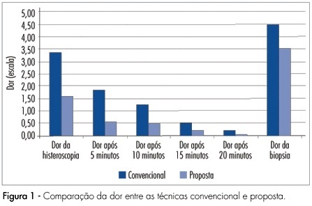
PURPOSE: to compare diagnostic hysteroscopy through vaginoscopy, using warm saline solution, with traditional technique, regarding to pain, patient satisfaction and feasibility of the procedure. METHODS: randomized clinical trial, involving 184 women, referred for diagnostic hysteroscopy, between May and December of 2006. Participants were randomized to be submitted to hysteroscopy by the proposed technique, which consisted of access through vaginoscopy using normal saline at 36ºC as distension medium, no speculum or cervical grasping, or by the traditional technique with CO2. In both techniques, a 2.7 mm hysteroscope was used. Pain was assessed by the analogical visual scale, during the procedure and every five minutes after it. RESULTS: the mean pain score was 1.60 in the proposed technique and 3.39 in the traditional technique (p<0.01). Lower pain scores were also observed after 5, 10 and 15 minutes (p<0,01) as well as after 20 minutes (p=0.056). In the proposed technique, 82.4% of the procedures were feasible, while, in the traditional technique, 84.9% were so (p=0.64). Satisfaction with the procedure was referred by 88.7% of women submitted to the proposed technique and by 76.3% of women submitted to the traditional technique (p<0.05). CONCLUSIONS: diagnostic hysteroscopy by the proposed technique resulted in less pain, same feasibility and greater satisfaction of patients.

Summary
Revista Brasileira de Ginecologia e Obstetrícia. 2007;29(6):285-290
DOI 10.1590/S0100-72032007000600002
PURPOSE: to evaluate the spreading of endometrial cells to the peritoneal cavity during diagnostic hysteroscopy. METHODS: a prospective, descriptive study involving 76 patients divided in two groups: one with 61 patients without malignant endometrial cancer, and the other with 15 patients with endometrial cancer. Two samples of peritoneal fluid were collected, one before (PF-1) and the other immediately after (PF-2) the diagnostic hysteroscopy. Spread to the peritoneal cavity was defined by the presence of endometrial cells in PF-2, with the absence of such cells in PF-1. The 5 mm diameter Storz’s hysteroscopy was used. Distention was obtained by CO2 with electronically controlled flow pressure of 80 mmHg. The PF was fixated in absolute alcohol (ratio1:1). The PF samples were centrifuged and aliquots were smeared and stained using the Papanicolaou method. Analyses were performed by the same observer. RESULTS: during the study, four patients (5.26%) were excluded for presenting endometrial cells in PF-1. In the remaining 72 patients, there was no spread of cells to the peritoneal cavity. In the non-endometrial cancer group, 88.1% (52/59) presented secretory endometrial phase, with correlation of 80% between the hysteroscopy and the biopsy. In the group with endometrial cancer, most of the patients were in stage I (92.3%). There was a 100% correlation between the hysteroscopy/biopsy and histopathology of the surgical sample. CONCLUSIONS: the diagnostic hysteroscopy with CO2 at flow pressure of 80 mmHg did not cause spread of endometrial cells to the peritoneal cavity in both groups, thus suggesting that the diagnostic hysteroscopy is safe for patients at high risk for endometrial cancer.
Summary
Revista Brasileira de Ginecologia e Obstetrícia. 2007;29(4):181-185
DOI 10.1590/S0100-72032007000400003
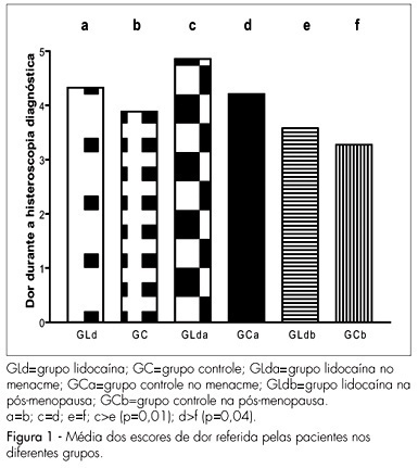
PURPOSE: to determine the efficacy of 10% lidocaine spray applied to the cervix before the procedure of diagnostic hysteroscopy, in order to reduce the painful process and the discomfort caused by the exam. METHODS: a total of 261 consecutive patients participated in the study, which was conducted from March 2004 to March 2005. The patients were randomly assigned to one of two groups: one group receiving topical lidocaine spray (lidocaine group - LdG) and the other, receiving no medication before the procedure (control group - CG). In the LdG patients, thirty milligrams of 10% lidocaine spray were applied to the surface of the cervix five minutes before hysteroscopy started. Immediately, after the end of the procedure, the patients from both groups were asked to respond to a questionnaire about pain and to quantify the pain, in centimeters, using a 10-cm non-graduated visual analog scale. The unpaired t test, the Mann-Whitney test and the chi2 test were used for statistical analyses, considering p significant if lower than 0.05. RESULTS: there was no statistically significant difference between groups regarding age, parity or percentage of patients in menacme or menopause, or regarding the indications for the procedure and the hysteroscopic findings. A biopsy was necessary in 57 of the 132 LdG patients and in 48 of the 129 CG patients (p=0.96). The mean pain score was 4.3±2.9 in LdG and 3.9±2.5 in CG (p=0.2). A difference in the mean pain score was observed only among patients in menacme and menopause receiving or not the lidocaine spray, with p=0.01 and p=0.04 respectively. CONCLUSIONS: the use of lidocaine spray during diagnostic hysteroscopy does not minimize the discomfort and pain of the patients and therefore should not be applied.

Summary
Revista Brasileira de Ginecologia e Obstetrícia. 1998;20(7):405-410
DOI 10.1590/S0100-72031998000700006
Objective: to demonstrate the effectiveness of video-hysteroscopic endometrial resection in the treatment of abnormal uterine bleeding. Patients and method: The authors studied 60 records of patients with abnormal uterine bleeding who did not respond to clinical treatment. Results: eighty-eight percent of the patients had adequate response to the treatment (53% oligomenorrhea and 35% amenorrhea). The complication rate was 8.3% (5 uterine perforations). Conclusion: video-hysteroscopic endometrial resection is an effective technique to treat abnormal uterine bleeding which failed to respond to clinical management. The intra and postoperative complication rates are low.
Summary
Revista Brasileira de Ginecologia e Obstetrícia. 2000;22(4):235-238
DOI 10.1590/S0100-72032000000400008
Purpose: to evaluate thermal balloon endometrial ablation in the management of menorrhagia. Study design: twenty patients were submitted to endometrial ablation using the thermal balloon device, between June 1996 and June 1997. Local anesthesia was used in 16 patients. The device was introduced into the uterine cavity. The duration of the procedure was 8 minutes and 30 seconds. Results: two patients (10%) did not show improvement of the symptons. Eighteen patients (90%) referred improvement of symptoms. There was no complication during and after the procedure. Conclusions: The thermal balloon seems to be safe and efficient in the management of menorrhagia.
Summary
Revista Brasileira de Ginecologia e Obstetrícia. 2004;26(7):527-533
DOI 10.1590/S0100-72032004000700004
PURPOSE: to evaluate the diagnostic accuracy of hysterosalpingography (HSG) and transvaginal sonography (TVS) in terms of detecting uterovaginal anomalies in women with a history of recurrent miscarriage. METHODS: eighty patients who presented two or more consecutive miscarriages were submitted to HSG, TVS and hysteroscopy (HSC). The following diagnoses were considered separately: uterine malformations, intrauterine adhesions and polypoid lesions. Hysteroscopy was the gold standard. The matching among the different methods was evaluated by the kappa coefficient and its significance was tested. The significance level was 0.05 (alpha=5%). Sensitivity, specificity, positive and negative predictive values, with 95% of statistical confidence interval, were calculated. RESULTS: uterovaginal anomalies were detected in 29 (36.3%) patients: 11 (13.7%) were uterine malformations, 17 (21.3%) intrauterine adhesions and one (1.3%) a polypoid lesion. The global matching between HSG and HSC was 85.5%, while between TVS and HSC it was only 78.7%. The best accuracy of HSG appeared to be for the diagnosis of uterine malformations and intrauterine adhesions (diagnostic accuracy of 97.5 and 95%, respectively). For the diagnosis of polypoid lesions, HSG had a diagnostic accuracy of only 92.5%, due to the low rate of positive predictive value (14.3%). TVS had a worse accuracy for all diagnoses, 93.7% for the diagnosis of uterine malformations and 85% for intrauterine adhesions, due to low sensitivity. CONCLUSIONS: histerosalpingography showed a good diagnostic accuracy for the diagnosis of uterine cavity diseases. TVS had good specificity, but with low sensitivity.
Summary
Revista Brasileira de Ginecologia e Obstetrícia. 2004;26(4):305-309
DOI 10.1590/S0100-72032004000400007
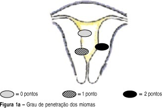
OBJECTIVE: to develop a new preoperative classification of submucous myomas to evaluate the viability and the degree of difficulty of hysteroscopic myomectomy. METHODS: forty-four patients were submitted to hysteroscopic resection of submucous myomas. The possibility of total resection of the myoma, the surgery duration, the fluid deficit, and the incidence of complications were evaluated. The myomas were classified by the Classification of the European Society of Endoscopic Surgery (CESES) and by the classification proposed (CP) by our group, that besides the degree of penetration of the myoma in the myometrium, adds the parameters: extent of the base of the myoma as related to the uterine wall, the size of the myoma in centimeters and its topography at the uterine cavity. For statistical analysis the Fisher test, the Student t test and the analysis of variance were used. Statistic significance was considered when the p-value was smaller than 0.05 in the bicaudal test. RESULTS: in 47 myomas the hysteroscopic surgery was considered complete. There was no significant difference among the three levels (0, 1 and 2) by CESES. By CP, the difference among the number of complete surgeries was significant (p=0.001) between the two levels (groups I and II). The difference between the surgery duration was significant when the two classifications were compared. In relation to the fluid deficit, just CP presented significant differences among the levels (p=0,02). CONCLUSIONS: the proposed classification includes more clues about the difficulties of the hysteroscopic myomectomy than the standard classification. It should be noted that the number of hysteroscopic myomectomies used for that analysis was modest, being interesting to evaluate the performance of the proposed classification in larger series of cases.
