Summary
Revista Brasileira de Ginecologia e Obstetrícia. 2008;30(4):190-195
DOI 10.1590/S0100-72032008000400006
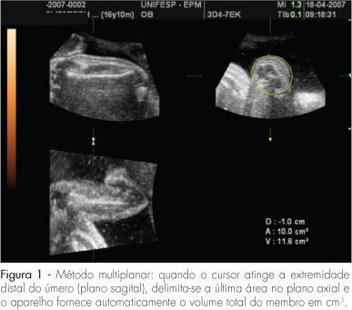
PURPOSE: to evaluate the accuracy of fetal upper arm volume, using three-dimensional ultrasound (3DUS), in the prediction of birth weight. METHODS: this prospective cross-sectional study involved 25 pregnancies without structural or chromosomal anomalies. Bidimensional parameters (biparietal diameter, abdominal circumference and femur length) and the 3DUS fetal upper arm volume were obtained in the last 48 hours before delivery. The multiplanar method, using multiple sequential planes with 5.0-mm intervals, was used to calculate fetal upper arm volume. Polynomial regressions were used to determine the best equation in the prediction of fetal weight. The accuracy of this new formula was compared with Shepard's and Hadlock's formulas. RESULTS: fetal upper arm volume was strongly correlated to birth weight (r=0.83; p<0.005). Linear regression was the best equation [birth weight=681.59 + 43.23 x fetal upper arm volume]. The fetal upper arm volume mean error (0 g), mean absolute error (196.6 g) and mean percent absolute error (6.5%) were lower than using Shepard's formula; however, the difference did not reach significance (p>0.05). Birth weight predicted by fetal upper arm volume had a mean error lower than Hadlock's formula, but this difference was not statistically significant (p>0.05). CONCLUSIONS: the accuracy of fetal upper arm volume obtained through 3DUS is similar to the accuracy of bidimensional ultrasound in the prediction of birth weight. These findings need to be confirmed by larger studies.
Summary
Revista Brasileira de Ginecologia e Obstetrícia. 2008;30(1):5-11
DOI 10.1590/S0100-72032008000100002
PURPOSE: to study the value of Doppler velocimetry of the ductus venosus, between the 11th and 14th weeks of pregnancy, associated to the nuchal translucency thickness measurement, in the detection of adverse fetal outcome. METHODS: a transversal and prospective study in which a total of 1,268 fetuses were studied consecutively. In 56 cases, a cytogenetic study was performed on material obtained from a biopsy of the chorionic villus and, in 1,181 cases, the postnatal phenotype was used as a basis for the result. In addition to the routine ultrasonographic examination, all the fetuses were submitted to measurement of the nuchal translucency thickness and to Doppler velocimetry of the ductus venosus. Aiming at prevalence and accuracy indices, sensitivity, specificity, positive predictive value, negative predictive value, probability of false-positive, probability of false-negative, reason of positive probability and reason of negative probability were calculated and analyzed. RESULTS: from the total of 1,268 fetuses, 1,183 cases were selected for analysis. From this number, 1,170 fetuses were normal (98.9%) and 13 fetuses presented adverse outcome at birth (1.1%), including fetal death (trisomy 21 and 22) in two cases; genetic syndrome (Nooman) in one case; two cases of polymalformed fetuses; cardiopathy in three cases; and other structural defects in five cases. The prevalence of the modified ductus venosus (wave A zero/reverse) in the studied population was of 14 cases (1.2%), with a false-positive rate of 0.7%. CONCLUSIONS: there is a significant correlation between the alteration of the ductus venosus Doppler velocimetry and the thickness of the nuchal translucency as an ultrasonographic marker for the first trimester of gestation, in the detection of adverse fetal outcome, especially serious malformations. The ductus venosus was able to diminish the false-positive result in comparison to the isolated use of the nuchal translucency thickness, improving considerably the positive predictive value of the test.
Summary
Revista Brasileira de Ginecologia e Obstetrícia. 2007;29(12):614-618
DOI 10.1590/S0100-72032007001200003
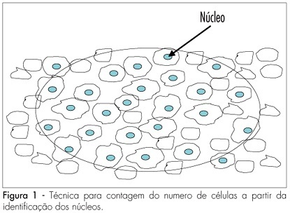
PURPOSE: to determine the variation of the number of ovarian follicles during fetal life. METHODS: twelve ovaries donated for research were included in our study, nine from fetuses and three from newborn babies who died in the first hour after being delivered with 39 weeks of pregnancy. Fetal age was confirmed both by the last menstrual period of the woman and by ultrasonography. Ovaries were fixed in formaldehyde, included in paraffin and serially sliced at 7 mm. At every 50 cuts, the obtained material was haematoxilin-eosin stained and evaluated with an optical microscope (400 X). The follicles were counted in ten different regions of the ovarian cortex, each region with an area of 625 mm². The presence of a nucleus was considered the parameter for counting. Follicular density, per 1 mm³ was calculated using the formula Nt=(No x St x t)/do, where Nt is the number of follicles; No is the mean number of follicles in 1 mm²; St is the total number of slices in 1 mm³; t is the slice thickness and do is the nuclei mean diameter. RESULTS: the gestational age of fetuses ranged from 24 to 39 weeks. The number of follicles per 0.25 mm² ranged from 10.9 ± 4.8 in a newborn to 34.7 ± 10.6 in another newborn. Among the fetuses, the least value was obtained in a 36 week-old fetus (11.1 ± 6.2) and the highest in a 28 week-old fetus (25.3 ± 9.6). The total number of slices per ovary ranged from six to 13, corresponding to follicles counted in areas from 15 to 32.5 mm². The total number of follicles ranged from 500,000 at the age of 22 weeks to > 1,000,000 at the age of 39 weeks. CONCLUSIONS: our results demonstrate different (increasing) densities of ovarian follicles along the gestational period, providing more knowledge about this still not well-known subject.
Summary
Revista Brasileira de Ginecologia e Obstetrícia. 2007;29(7):352-357
DOI 10.1590/S0100-72032007000700005
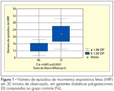
PURPOSE: to analyze the pattern of fetal breathing movements (FBM) in diabetic pregnant women in the third trimester of pregnancy. METHODS: sixteen pregestational diabetic and 16 nondiabetic (control group) pregnant subjects were included fulfilling the following criteria: singleton, between 36-40 weeks of gestation, absence of other maternal diseases and absence of fetal anomalies. The fetal biophysical profile (FBP) was performed to evaluate the following parameters: fetal heart rate, FBM, fetal body movements, fetal tone and amniotic fluid index. The FBM was evaluated for 30 minutes, period when the examination was integrally recorded in VHS video for posterior analysis of the number of FBM episodes, the duration of each episode and the fetal breathing movements index (BMI). The BMI was calculated by the formula: (interval of time with FBM/total time of observation) x 100. At the beginning and in the end of the FBP maternal glucose levels were checked. The results were analyzed by the Mann-Whitney U-test and the Fisher exact test, adopting a level of significance of 5%. RESULTS: the glucose levels demonstrated significantly superior average in the diabetic group (113.3±35.3 g/dL) in relation to the normal group (78.2±14.8 g/dL, p<0.001). The average of the amniotic fluid index was higher in the group of the diabetic cases (15.5±6.4 cm) when compared with controls (10.6±2.0 cm; p=0.01). The average of the number of FBM episodes was superior in the diabetic ones (22.6±4.4) in relation to controls (14.8±2.3; p<0.0001). The average of the BMI in the diabetic patients (54.6±14.8%) was significantly higher than that in the control group (30.5±7.4%, p<0.0001). CONCLUSIONS: the elevated blood glucose levels can be associated with a different pattern in the FBM of diabetic mothers. The use of this parameter of the FBP, in the obstetric practice, must be considered with concern in diabetic pregnancies.
Summary
Revista Brasileira de Ginecologia e Obstetrícia. 2007;29(4):192-199
DOI 10.1590/S0100-72032007000400005
PURPOSE: to evaluate and compare the knowledge and the opinion of gynecologists and obstetricians regarding termination of pregnancy, in 2003 and 2005. METHODS: a structured and pre-tested questionnaire was sent to all the members of the Brazilian Federation of Gynecologists and Obstetricians (FEBRASGO). They were asked to answer the questions, anonymously, and return the questionnaire in a stamped envelope provided. They were asked about their knowledge of and opinion on Brazilian legislation related to abortion. RESULTS: in both surveys the percentage of doctors who knew under which circumstances abortion was not penalized was over 80%. However, there was a significant reduction in the percentage of doctors who knew that abortion was legal if the woman’s life was at risk. The participants who knew that abortion because of a severe congenital malformation of the fetus was not currently permitted by law increased by a third. The percentage of doctors in favor of allowing abortion increased consistently for the various circumstances presented. The proportion of those who thought that abortion should not be permitted in any circumstances decreased. The percentage of those who judged that the legal consents should not be modified decreased. There was an increase in the proportion of those who considered that abortion should not be considered a crime under any circumstance. CONCLUSIONS: in general, it seems that people have been thinking more about induced abortion during the time elapsed between the two surveys. Nevertheless, there is the need to correctly inform Brazilian gynecologists and obstetricians on the laws and norms that regulate the practice of legal abortion in the country, so as to ensure that women who need one have, in fact, access to this right.
Summary
Revista Brasileira de Ginecologia e Obstetrícia. 1998;20(5):237-243
DOI 10.1590/S0100-72031998000500002
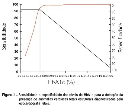
We analyze prospectively the existence of a relationship between the mother's glycemic control, in the first half of pregnancy, and the occurrence of abnormal fetal cardiac abnormalities, in pregnant women with diabetes mellitus. In 127 pregnant women, the level of glycosylated hemoglobin was determined on the first visit during prenatal care. Nine patients had type I diabetes, 77 type II and 41 gestational diabetes mellitus (GDM). All mothers were submitted to detailed fetal echocardiography, during the 28th ± 4.127 week of gestation. In 31 (24.4%) of the 127 fetuses cardiac anomalies were detected. In 10 (7.87%) an isolated cardiac anomaly was identified. Mean HbA1c in the group of pregnant women without cardiac anomalies (5.64%) was statistically different from the group with anomalies (10.14%) (p<0.0001). The receiver-operator characteristic, representing the balance between sensitivity (92.83%) and specificity (98.92%) in the diagnosis of structural cardiac abnormalities, showed a cut-off point at the 7.5% HbA1c level. In nine of ten fetuses with structural cardiac anomalies, the maternal level of HbA1c was higher than 7.5%. The difference between means of the groups with and without myocardial hypertrophy diagnosed as isolated anomaly (MCHP) was not statistically significant, when considering both type II diabetes and GDM subgroups. In conclusion, levels of HbA1c higher than 7.5% were associated with most cases of echocardiographycally diagnosed structural cardiac anomalies. On the other hand, this test was not useful to discriminate conceptus with MCHP.
Summary
Revista Brasileira de Ginecologia e Obstetrícia. 1998;20(9):517-524
DOI 10.1590/S0100-72031998000900005
Purpose: to determine the behavior of doppler velocimetry during the course of risk pregnancies and to compare the perinatal results obtained for concepti with retarded intrauterine growth (RIUG) with those for concepti considered adequate for gestational age (AGA). Methods: a prospective study of the evolution of doppler ultrasound was made in 38 pregnant women with of idiopathic intrauterine growth retardation (IUGR) in previous pregnancy. A relationship was established between this antecedent and the new pregnancy. The pregnant women studied were divided into two groups in agreement with their neonates birthweight. Group 1 was associated with IUGR and group 2 with adequate birth weight. IUGR was confirmed in 23.7% of the cases. Umbilical and uterine artery doppler velocimetry was performed from 20 to 40 weeks of gestation. Middle cerebral artery doppler velocimetry was analyzed after 28 weeks of gestation, twice a month, being the last valued examination before birth. Results: the uterine and umbilical artery ratio at 24 and 28 weeks of gestation, respectively, correlated with the presence of IUGR. There was no difference between the two groups regarding the presence or absence of a small notch in the uterine artery wave form and middle cerebral artery doppler velocimetry ratio, at the last examination before birth. There was a relationship between neonatal stay in hospital for more than three days and the presence of IUGR. Conclusions: doppler ultrasound should be used in the follow-up of cases with a high risk of IUGR. It allows the detection of the fetuses at high risk of hypoxia and, by interrupting the pregnancy, fetal distress-related complications may be avoided.
Summary
Revista Brasileira de Ginecologia e Obstetrícia. 1998;20(8):443-448
DOI 10.1590/S0100-72031998000800003
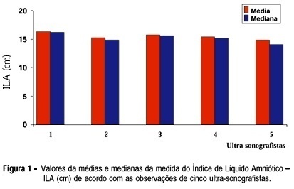
Purpose: to demonstrate the interobserver variation existing in the ultrasonographic measurement of amniotic fluid index (AFI) and in the measurement of pocket area, and to compare these two methods. In addition, an attempt was made to establish the intraobserver variation in the measurement of this index. Methods: values of AFI, described by Phelan et al.18, were studied in a group of 80 pregnant women considered to be clinically normal, seen at the Ultrasonography and Medical Updating School of Ribeirão Preto and in the Department of Gynecology and Obstetrics of the Faculty of Medicine of Ribeirão Preto, University of São Paulo (FMRP-USP). All pregnant women had a gestational age of more than 24 weeks. Fifty of these patients were submitted to AFI evaluation by 5 different ultrasonographists using the same equipment and during the same period of time, in order to determine the interobserver variation of this index. In addition, planimetric measurement of the area was performed by 2 of these 5 ultrasonographists, selected at random, in an attempt to determine interobserver variation in area measurement. Another group of 30 pregnant women was evaluated by the same ultrasonographist in an attempt to evaluate intraobserver variation in terms of AFI measurement. Results: There was a significant interobserver variation in AFI measurement and a significant variation in area measurement. However, the intraobserver variation in AFI measurement was nonsignificant. There was a correlation between AFI and area measurements. Conclusions: we emphasize the obstetrical applicability of this index and the easier execution of this method compared to area measurement, despite the importance of both procedures.
