Summary
Revista Brasileira de Ginecologia e Obstetrícia. 2013;35(3):123-129
DOI 10.1590/S0100-72032013000300006
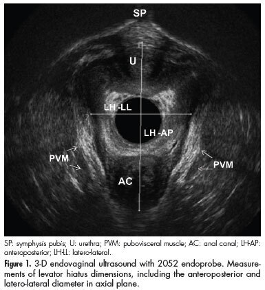
PURPOSE: To determine anatomical and functional pelvic floor measurements performed with three-dimensional (3-D) endovaginal ultrasonography in asymptomatic nulliparous women without dysfunctions detected in previous dynamic 3-D anorectal ultrasonography (echo defecography) and to demonstrate the interobserver reliability of these measurements. METHODS: Asymptomatic nulliparous volunteers were submitted to echo defecography to identify dynamic dysfunctions, including anatomical (rectocele, intussusceptions, entero/sigmoidocele and perineal descent) and functional changes (non-relaxation or paradoxical contraction of the puborectalis muscle) in the posterior compartment and assessed with regard to the biometric index of levator hiatus, pubovisceral muscle thickness, urethral length, anorectal angle, anorectal junction position and bladder neck position with the 3-D endovaginal ultrasonography. All measurements were compared at rest and during the Valsalva maneuver, and perineal and bladder neck descent was determined. The level of interobserver agreement was evaluated for all measurements. RESULTS: A total of 34 volunteers were assessed by echo defecography and by 3-D endovaginal ultrasonography. Out of these, 20 subjects met the inclusion criteria. The 14 excluded subjects were found to have posterior dynamic dysfunctions. During the Valsalva maneuver, the hiatal area was significantly larger, the urethra was significantly shorter and the anorectal angle was greater. Measurements at rest and during the Valsalva maneuver differed significantly with regard to anorectal junction and bladder neck position. The mean values for normal perineal descent and bladder neck descent were 0.6 cm and 0.5 cm above the symphysis pubis, respectively. The intraclass correlation coefficient ranged from 0.62-0.93. CONCLUSIONS: Functional biometric indexes, normal perineal descent and bladder neck descent values were determined for young asymptomatic nulliparous women with the 3-D endovaginal ultrasonography. The method was found to be reliable to measure pelvic floor structures at rest and during Valsalva, and might therefore be suitable for identifying dysfunctions in symptomatic patients.

Summary
Revista Brasileira de Ginecologia e Obstetrícia. 2012;34(4):158-163
DOI 10.1590/S0100-72032012000400004
PURPOSE: To verify cervical length using transvaginal ultrasonography in pregnant women between 28 and 34 weeks of gestation, correlating it with the latent period and the risk of maternal and neonatal infections. METHODS: 39 pregnant women were evaluated and divided into groups based on their cervical length, using 15, 20 and 25 mm as cut-off points. The latency periods evaluated were three and seven days. Included were pregnant women with live fetuses and gestational age between 28 and 34 weeks, with a confirmed diagnosis on admission of premature rupture of membranes. Patients with chorioamnionitis, multiple gestation, fetal abnormalities, uterine malformations (bicornus septate and didelphic uterus), history of previous surgery on the cervix (conization and cerclage) and cervical dilation greater than 2 cm in nulliparous women and 3 cm in multiparae were excluded from the study. RESULTS: A <15 mm cervical length was found to be highly related to a latency period of up to 72 hours (p=0.008). A <20 mm cervical length was also associated with a less than 72 hour latency period (p=0.04). A <25 mm cervical length was not found to be statistically associated with a 72 hour latency period (p=0,12). There was also no significant correlation between cervical length and latency period and maternal and neonatal infection. CONCLUSION: The presence of a short cervix (<15 mm) was found to be related to a latency period of less than 72 hours, but not to maternal or neonatal infections.
Summary
Revista Brasileira de Ginecologia e Obstetrícia. 2011;33(9):258-263
DOI 10.1590/S0100-72032011000900007
PURPOSE: To evaluate the coverage of Pap smear cytology at Basic Family Health Units (BFHU) and to describe the characteristics of non-performance of this test in the last three years. METHODS: A cross-sectional study was conducted in Rio Grande (RS), Brazil, in areas covered by the Family Health Teams Family (FHT). The interviews were conducted by students participating in the Health-PET, at women’s home. Crude analysis was performed using SPSS software to calculate prevalence ratio, 95% confidence intervals and p value. Multivariate analysis was performed by Poisson regression using Stata 9.0 software, which were included the variables with p value of up to 0.20 in the crude analysis. At the first level, the variables were age, having a partner, and literacy. At the second level, the variables were number of visits and offer of a Pap smear. RESULTS: The prevalence of Pap cytology performed 36 months ago or less was 66.3%. In adjusted analysis, women aged 19 years or less (p<0.001), without a partner (p<0.001), illiterate (p= 0.01), who had never consulted at the basic unit (p=0.02) and who had not been offered the examination during the visit (p=0.006), were more likely not to have had a cytopathology exam in the last 36 months. CONCLUSION: The local health proved to be ineffective and inequitable. Ineffective because it covers fewer women than indicated by the World Health Organization and uneven because access to this test varied according to some characteristics of the users.
Summary
Revista Brasileira de Ginecologia e Obstetrícia. 2010;32(8):386-392
DOI 10.1590/S0100-72032010000800005
PURPOSE: to evaluate the incidence and direct economic impact of cervical cancer (CC) in Roraima, in 2009, and to analyze the epidemiological profile of patients with this disease. METHODS: the histopathologic reports issued in Roraima in 2009 were reviewed, as were hospital records of female patients under treatment for cancer. Clinical data and medical procedures related to CC were recorded. CC carriers were treated under expenses of the public Brazilian health system (SUS) in Roraima underwent an interview dealing with socio-economic topics. RESULTS: we registered 90 cases of CC and high grade pre-invasive lesions. Roraima has the highest incidence of CC of Brazil (46.21 cases/100,000 women), which is 3 times higher than that of breast cancer, comparable to low-income developing countries. The epidemiological profile shows patients with economic deprivation, social disadvantage, low education, early first intercourse (mean age is 13.8 years), and high parity (medium of 5.5 gestations). Among the patients included in this report, 71.7% had never been submited to a Pap smear, and ignorance about it was the main reported reason (47.4%). As a public health problem, the management of CC generates direct annual expenditures of more than R$ 600,000, with an average cost per patient of R$ 8,711. CONCLUSIONS: CC is the most common cancer among women from Roraima, and represents a serious public health problem in Roraima. Its high economic impact favors the implementation of preventive strategies from the standpoint of cost-effectiveness. The profile of patients reveals the ineffectiveness of preventive services in reaching patients with a socio-economic exclusion profile at high risk for cervical cancer.
Summary
Revista Brasileira de Ginecologia e Obstetrícia. 2010;32(8):368-373
DOI 10.1590/S0100-72032010000800002
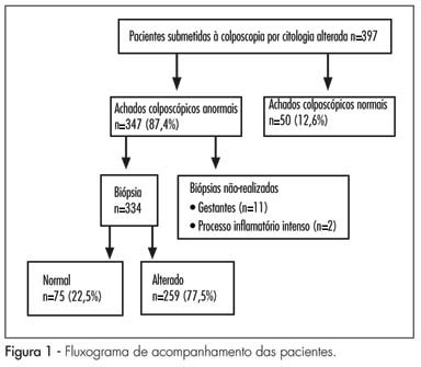
PURPOSE: to evaluate the agreement between conventional cytology using the Papanicolaou test, repeated at the time of colposcopy, with colposcopic and histopathological findings. METHODS: the study was carried out at the central public health laboratory of the state of Pernambuco between January and July, 2008, involving 397 women referred for colposcopic evaluation following an abnormal cervical smear test. Cytology was repeated at the time of colposcopy using conventional method, with particular attention being paid to the presence of abnormal colposcopic findings. The nomenclature used for cytology was the 2001 Bethesda system terminology, while that used for histology was the World Health Organization 1994 classification. Cytology performed at the time of colposcopy was compared with colposcopy and with histopathology obtained by colposcopy-directed biopsy. The Kappa coefficient was used to evaluate the agreement between methods, as well as the χ2 test, with the level of significance set at 5%. RESULTS: poor agreement was found between cytology performed at the time of colposcopy and colposcopic findings (K=0.33; 95%CI=0.21-0.45) and between colposcopy and histopathology (K=0.35; 95%CI=0.39-0.51). Cytology performed at the time of colposcopy compared with histopathology revealed a Kappa of 0.41 (95%CI=0.29-0.530), which was considered to reflect moderate agreement. CONCLUSIONS: agreement was better between cytology and histopathology than between colposcopy and cytology or between colposcopy and histopathology.

Summary
Revista Brasileira de Ginecologia e Obstetrícia. 2010;32(6):286-292
DOI 10.1590/S0100-72032010000600006
PURPOSE: to evaluate the expression of E-cadherin in cervical lesions of patients suffering from HIV infection. METHODS: we conducted a study with 77 patients with cervical HPV infection, 40 of them were HIV seropositive and 37 HIV seronegative who underwent colposcopy and a biopsy of the cervix. The material obtained by biopsy of the cervix was sent for histopathologic and immunohistochemical study. Sections were obtained and mounted on silanized slides and examined by an observer who was blind to patient serology. E-cadherin antibody, clone NHC-38 diluted 1:400 (DAKO) and the Novolink polymer system (Novocastra) were used. The expression of E-cadherin was determined on the epithelial cell membrane based on the extent of the stained area. The χ2 test with Yates correction or the Fisher's Exact test was used for comparison of the proportion in univariate analysis. All the variables with p<0.25 were included in the logistic regression model, called initial model. The analyses were carried out using the SPSS software, with the level of significance set at 5%. RESULTS: the expression of E-cadherin was observed in up to the internal 1/3 of the epithelium in 59.3% of cases and in up to 2/3 of the epithelium in 11.1% of cases, but in 29.6% of cases the expression was identified throughout the thickness of the epithelium in HIV-seronegative patients. In contrast, in HIV-seropositive patients, 45.9% showed expression up to 1/3 of the epithelium, 13.5% showed expression in up to 2/3 of the epithelium, and 40.5% showed expression throughout the thickness of the epithelium. E-cadherin expression did not differ between groups (p=0.5). However, the multivariate analysis identified a significant association between high-grade cervical injury and E-cadherin expression in 2/3 and 3/3 of the epithelium (p=0.001; χ2=36.9). CONCLUSIONS: the expression of E-cadherin in the epithelial cell membrane is not associated with infection by the human immunodeficiency virus, but with the degree of intraepithelial cervical injury.

Summary
Revista Brasileira de Ginecologia e Obstetrícia. 2009;31(4):203-209
DOI 10.1590/S0100-72032009000400008
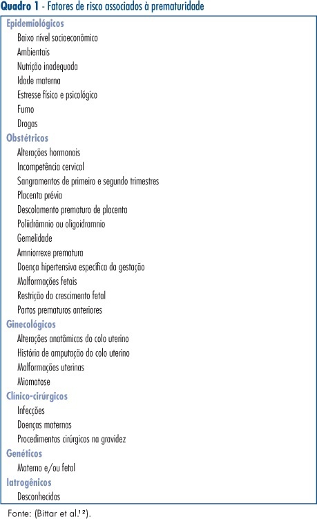
Among the clinical factors for preterm birth, some confer substantial increased risk, including a history of preterm birth, multiple gestation and vaginal bleeding in the second trimester. However, these factors are present only in a minority of women who ultimately deliver preterm and thus have low sensitivity. Cervical dilatation, effacement and position as determined by manual examination have been related to an increased risk of preterm birth but also suffer from low sensitivity and positive predictive values. Cervical length measured with transvaginal ultrasound has also been related to an increased risk of preterm birth as cervical length decreases. The reported sensitivity is better than other tests, but positive predictive value is low. The principal utility of the fetal fibronectin assay lies in its negative predictive value in symptomatic women. Increased sensitivity has been reported when cervical length is used in combination with fetal fibronectin.

Summary
Revista Brasileira de Ginecologia e Obstetrícia. 2008;30(9):437-444
DOI 10.1590/S0100-72032008000900003
PURPOSE: to verify the accuracy of uterine cervix cytology for HPV diagnosis, as compared to polymerase chain reaction (PCR) in samples of women with HIV. METHODS: 158 patients who had undergone a first collection of material from the uterine cervix with Ayre's spatula for PCR were included in the study. Then, another collection with Ayre's spatula and brush for oncotic cytology was performed. Only 109 slides were reviewed, as 49 of them had already been destructed for have being filed for over two years. RESULTS: the prevalence of HPV was 11% in the cytological exam and 69.7% in the PCR. Age varied from 20 to 61 years old, median 35 years. The HIV contagious route was heterosexual in 91.8% of the cases, and 79.1% of the patients had had from one to five sexual partners along their lives. The most frequent complaint was pelvic mass (5.1%), and 75.3% of the women had looked for the service for a routine medical appointment. The categorical variable comparison was done through contingency tables, using the χ2 test with Yates's correction to compare the ratios. The Fisher's test was used when one of the expected rates was lower than five. In the comparison of diagnostic tests, sensitivity, specificity and similarity ratios have been calculated. Among the 76 patients with HPV, detected by PCR, only 12 had the diagnosis confirmed by cytology (sensitivity=15.8%), which on the other hand did not present any false-positive results (specificity=100%). Concerning the HPV presence, the cytological prediction for positive results was 100% and 33.3% for negative, when both results were compared. Among the 12 patients with HPV positive cytology, four (33.3%) presented cervical intraepithelial neoplasia (OR=56; positive similarity ratio=positive infinity; negative similarity ratio=0.83). CONCLUSIONS: As the cytology specificity is quite high, it is possible to rely on the positive result, which means that a positive result will surely indicate the presence of HPV. The low sensitivity of cytology does not qualify it as a survey exam for HPV detection in this female group.