Summary
Revista Brasileira de Ginecologia e Obstetrícia. 2005;27(9):534-540
DOI 10.1590/S0100-72032005000900006
PURPOSE: to evaluate the role of morphological (12) and Doppler velocimetry (17) ultrasonographic features, in the detection of lymph node metastases in breast cancer patients. METHODS: 179 women (181 axillary cavities) were included in the study from January to December 2004. The ultrasonographic examinations were performed with a real-time linear probe (Toshiba-Power Vision-6000 (model SSA-370A)). The morphological parameters were studied with a frequency of 7.5-12 MHz. A frequency of 5 MHz was used for the Doppler velocimetry parameters. Subsequently, the women were submitted to level I, II and III axillary dissection (158), or to the sentinel lymph node technique (23). Sensitivity, specificity, and positive and negative predictive values were calculated for each parameter. The decision tree test was used for parameter association. The cutoff points were established by the ROC curve. RESULTS: at least one lymph node was detected in 173 (96%) of the women by the ultrasonographic examinations. Histological examination detected lymph node metastases in 87 women (48%). The best sensitivity among the morphological paramenters was found with the volume (62%), the antero-posterior diameter (62%) and the fatty hilum placement (56%). Though the specificity of the extracapsular invasion (100%), border regularity (92%) and cortex echogenicity (99%) were high, the sensitivity of these features was too low. None of the Doppler velocimetry parameters reached 50% sensitivity. The decision tree test selected the ultrasonographic parametners: fatty hilum placement, border regularity and cortex echogenicity, as the best parameter association. CONCLUSION: the detection of axillary cavity lymph node stage by a noninvasive method still remains an unfulfilled goal in the treatment of patients with breast cancer.

Summary
Revista Brasileira de Ginecologia e Obstetrícia. 2005;27(8):473-478
DOI 10.1590/S0100-72032005000800007
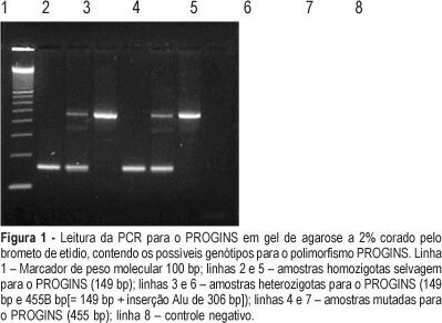
PURPOSE: to analyze the correlation between PROGINS polymorphism and breast cancer. METHODS: a case-control study was carried out from April to October 2004. The genotypes of 50 women with breast cancer and 49 healthy women were analyzed. The 306-base pair Alu insertion polymorphism in the G intron of progesterone receptor gene was detected by polymerase chain reaction and analyzed on 2% agarose gel stained with ethidium bromide. The control and experimental groups were compared regarding genotypes using the statistical Epi-Info 6.0 program and for frequencies the exact Fisher test or chi2 test were used. p value smaller p than 5% was considered to be significant. RESULTS: in relation to PROGINS we found in the studied population a prevalence of 62 (62.6%) wild homozygous, 35 (35.3%) heterozygous individuals and two (2.1%) cases with the presence of the mutation. Regarding PROGINS polymorphism, significance was not evidenced when cases and controls were compared, as related to homozygosis (62 vs 65.3%), heterozygosis (36 vs 34,6%) or the mutation (2.0 vs 2.1%), with p=0.920 (OR=1.01), 0.891 (OR=1.06), and 0.988 (OR=1.10), respectively. CONCLUSIONS: the results show that single-gene PROGINS polymorphism does not confer a substantial risk of breast cancer to its carriers.
Summary
Revista Brasileira de Ginecologia e Obstetrícia. 2005;27(7):415-420
DOI 10.1590/S0100-72032005000700008
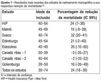
PURPOSE: to evaluate the cost of preventive mammographic screening in climacteric women, as compared to the cost of breast cancer treatment in more advanced stages. METHODS: one thousand and fourteen patients attended at the Climacteric outpatient service of the Gynecology Department, Federal University of São Paulo Paulista School of Medicine, were included in the study and submitted to mammographic test. All mammographic test's were analyzed by the same two physicians and classified according to the BI-RADS (Breast Imaging Reporting and Data System American College of Radiology) categories. The detected lesions were submitted to cytological and histological examination. RESULTS: the final diagnostic impression of the 1014 examinations, according to the classification of BI-RADS categories was: 1=261, 2=671, 3=59, 4=22 and 5=1. The invasive procedures were performed through a needle guided by ultrasound or stereotactic examinations: 33 fine-needle aspiration biopsies, 6 core biopsies guided by ultrasound and 20 core biopsies guided by stereotactic examination. Five cancer diagnoses were established. The total cost of this screening based on Brazilian procedure values was R$ 76,593.79 (25,534 dollars). Therefore, the cost of the diagnosis of the five cases of cancer in this screening was R$ 15,318.75 (5,106 dollars) each. However, the average cost per patient screened was R$ 75.53 (25 dollars). CONCLUSIONS: considering that the total treatment cost of only one case of breast cancer in advanced stage including hospital costs, surgery, chemotherapy, radiotherapy and hormonal treatment is similar to the cost of 1,000 mammographic screenings in climacteric women, it may be concluded that the cost of the early cancer diagnosis program is worth it and should be included in the public health program, as a way of lowering the public health expense.
Summary
Revista Brasileira de Ginecologia e Obstetrícia. 2005;27(6):340-346
DOI 10.1590/S0100-72032005000600008
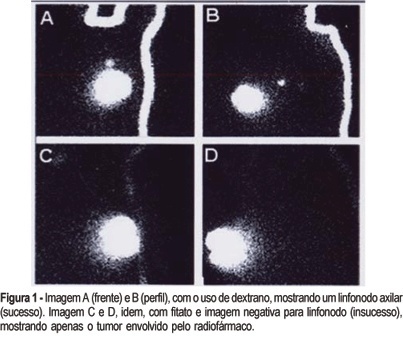
PURPOSE: a case-control study comparing two radiocolloids used in scintigraphy to map the sentinel lymph nodes (SLN) in breast cancer patients. METHODS: forty patients were prospectively enrolled between May 2002 and April 2004, after signing an informed consent form. In the present double-blind study, each patient was submitted twice to the same examination, a mammary scintigraphy, one with 99mTc-dextran 500 (dextran) and the other with 99mTc-phytate (phytate), on different days. A volume of 2 ml with 1-1.5 mCi of each radiopharmaceutical, in divided aliquots, was injected in the breast parenchyma in four points around in the tumor and the subcutaneous area superficial to the tumor. The image was obtained 2 h after the injection, using a gamma camera with high-resolution collimator. The lymph nodes were identified by anterior and lateral static scintigraphic images. Statistical analysis was done with the use of McNemar and Z tests. RESULTS: in the analysis of the 40 patients, we had 15 pairs with positive identical images, 4 pairs with negative images and 21 pairs with inconsistent images, either because one of them was negative, or because the SLN numbers were different. When the protocol was opened, we found 35 and 27 positive images and 5 and 13 negative images for dextran and phytate treatment groups, respectively. Among the negative images, 4 were shared by both groups. The McNemar test, used for the statistical analysis, showed p=0.026, odds ratio (OR) = 0.11 with 95% CI 0.01 < OR < 0.85. The accuracy, evaluated by the success ratio of the SLN mapping, was 67.5% for phytate and 87.5% for dextran, with p=0.032. Analysis of variance of the SLN number in lymphoscintigraphy images showed p=0.008. CONCLUSION: these results recommend the use of dextran instead of phytate for the SLN study of breast carcinoma by scintigraphy, when the same methodology is being used.
Summary
Revista Brasileira de Ginecologia e Obstetrícia. 2000;22(5):307-310
DOI 10.1590/S0100-72032000000500009
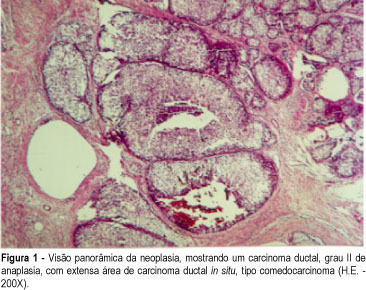
Polymastia is a usual problem in Mastology clinics and the possibility of cancer must be taken into consideration, as much as in any other mammary tissue. In the present study the case of a 48-year-old patient, submitted to the excision of the left axillary breast for cosmetic purposes is reported. The histological examination showed an invasive ductal carcinoma with an extensive in situ component. The patient was submitted to a wide excision plus axillary lymphadenectomy and radiation therapy. The frequency, diagnosis, prognosis and treatment of cancer in supernumerary breasts is also reviewed.
Summary
Revista Brasileira de Ginecologia e Obstetrícia. 2000;22(7):455-458
DOI 10.1590/S0100-72032000000700009
Primary angiosarcoma of the breast is a rare tumor, which appears between 14 and 82 years, with an average of 35 years of age. Its predominant clinical aspect is a painful mass with diffuse increase in the breast and violet or blackened color. Equally to other cases of sarcoma, the medium size of the lesion is approximately 5 cm at the diagnosis. Histologically, it is characterized by the proliferation of endothelial cells that form vascular channels linked to each other infiltrating glandular structures and fatty tissue. Its histological diagnosis is difficult and not always the right diagnosis is immediately established, mainly in the cases of a low malignancy degree, due to limited biopsy material. Because of the difficult diagnosis and aggressivity, it is a neoplasia with ominous prognosis, due to frequent metastasis. In our service, a 18-year-old patient presented with a painful lump which grew quickly. It was biopsied and a hemangioma was diagnosed, a wide excision being indicated. Three months later, she suffered a tumoral relapse, that was biopsied again and mastectomy was indicated, because it was an angiosarcoma with low degree of malignancy. After other relapses, chemotherapy was indicated and later, radiotherapy. During radiotherapy she developed new metastases, and died of pulmonary metastasis.
