Summary
Revista Brasileira de Ginecologia e Obstetrícia. 2012;34(6):278-284
DOI 10.1590/S0100-72032012000600007
PURPOSE: To evaluate the anatomical distribution of deep infiltrating endometriosis (DIE) lesions in a sample of women from the South of Brazil. METHODS: A prospective study was conducted on women undergoing surgical treatment for DIE from January 2010 to January 2012. The lesions were classified according to eight main locations, from least serious to worst: round ligament, anterior uterine serosa/vesicouterine peitoneal reflection, utero-sacral ligament, retrocervical area, vagina, bladder, intestine, ureter. The number and location of the DIE lesions were studied for each patient according to the above-mentioned criteria and also according to uni- or multifocality. The statistical analysis was performed using Statistica version 8.0. The values p<0.05 were considered statistically significant. RESULTS: During the study period, a total of 143 women presented 577 DIE lesions: uterosacral ligament (n=239; 41.4%), retrocervical (n=91; 15.7%), vagina (n=50; 8.7%), round ligament (n=50; 8,7%), vesico-uterine septum (n=41; 7.1%), bladder (n=12; 2.1%), and intestine (n=83; 14.4%), ureter (n=11; 1.9%). Multifocal disease was observed in the majority of patients (p<0.0001), and the mean number of DIE lesions per patient was 4. Ovarian endometrioma was present in 57 women (39.9%). Sixty-five patients (45.4%) presented intestinal infiltration on histological examination. A total of 83 DIE intestinal lesions were distributed as follows: appendix (n=7), cecum (n=1) and rectosigmoid (n=75). The mean number of intestinal lesions per patient was 1.3. CONCLUSIONS: DIE has a multifocal pattern of distribution, a fact of fundamental importance for the definition of the complete surgical treatment of the disease.
Summary
Revista Brasileira de Ginecologia e Obstetrícia. 2012;34(6):285-289
DOI 10.1590/S0100-72032012000600008
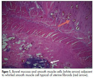
Extrauterine leiomyomas are rare, benign, and may arise in any anatomic sites. Their unusual growth pattern may even mimic malignancy and can result in a clinical dilemma. Occasionally, uterine leiomyomas become adherent to surrounding structures. They also develop an auxiliary blood supply, and lose their original attachment to the uterus, thus becoming 'parasitic'. Parasitic myomas may also be iatrogenically created after uterine fibroid surgery, particularly if morcellation is used. This report presented two cases of parasitic myomas with sepsis, both requiring right hemicolectomy. It reviewed the pertinent literature.
