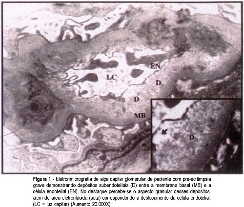
PURPOSE: to investigate the glomerular alterations in patients with severe preeclampsia, as well as to evaluate the evolution of these lesions, relating them to the moment of the renal biopsy. METHODS: seventy-two pregnant women with hypertensive syndrome underwent renal biopsy in the puerperium. Appropriate samples for electron microscopic examination were obtained from 39 patients and grouped as follows: 25 with preeclampsia and 14 with superimposed preeclampsia. Biopsy findings were classified into: normal kidney, endothelial cell edema, mesangial expansion, mesangial interposition, subendothelial fibrinoid deposits, and podocyte fusion. RESULTS: the most frequent alterations found in both groups were subendothelial fibrinoid deposits and podocyte fusion. Endothelial edema was present in 84% of the preeclampsia patients and in 92.9% of the superimposed preeclampsia cases. There was no association between the degree of hypertension and the severity of endothelial edema. A tendency to mesangial interposition was observed in patients who had a biopsy after the seventh day after delivery. Podocyte fusion showed a significant association with 24-hour proteinuria. CONCLUSIONS: the above mentioned glomerular alterations represent a spectrum of complex and dynamic lesions that together represent the ultrastructural characteristics of preeclampsia which should no longer be diagnosed based only on the presence or absence of endothelial edema.
Search
Search in:


Comments