Revista Brasileira de Ginecologia e Obstetrícia. 2015;37(4):149-151
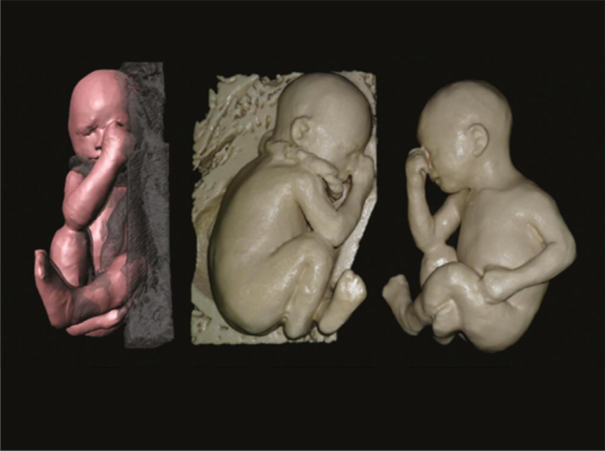
Improvements in the technology for non-invasive acquisition and visualization of images have brought about great advances in medicine, particularly in the diagnostic and prognostic assessments of foetal anomalies. In general, ultrasound (US) and magnetic resonance imaging (MRI) are two techniques that are used to acquire images of the foetus and uterus during pregnancy. The computed tomography (CT) also provides detailed foetal images, especially those of the foetal skeleton from the 30th week; however, its application is limited to the diagnosis of complex skeletal disorders because of the effects of ionizing radiation.
The earliest attempts to graphically represent the foetus date back to the 15thcentury A.D. Artistic renderings from that period onwards can be found in museums and private collections worldwide. Leonardo da Vinci is among the artists who refined the quality of the foetus visual representation. By means of several anatomical studies, da Vinci illustrated the entire process of foetal development.
[…]
Search
Search in:
Search
Search in:


Comments