Revista Brasileira de Ginecologia e Obstetrícia. 2004;26(1):59-64

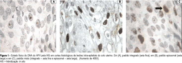
PURPOSE: to carry out a molecular study (in situ hybridization) on patients who present intraepithelial lesions of the uterine cervix, and to assess the frequency and the physical state of the human papillomavirus (HPV). METHODS: histological sections of biopsies of the uterine cervix from 84 patients were evaluated by in situ hybridization, with a broad-spectrum probe, which allows the identification of the HPV types 6, 11, 16, 18, 31, 33, 35, 39, 42, 45, and 56 and with specific probes for HPV types 6, 11, 16, 18, 31, and 33. The physical patterns of HPV DNA found were: episomal, when the entire nucleus stains with biotin (brown); integrated – one or two brown points in the hybridized nucleus, or mixed, associating both patterns. Of the 84 patients evaluated, 31 (36.9%) had low-grade squamous intraepithelial lesions (LSIL), and 53 (63.1%) had high-grade squamous intraepithelial lesions (HSIL) on histological examination. Fisher’s exact test was used for the statistical analysis. RESULTS: considering all the cases, 46 (54.7%) were positive for HPV DNA with the broad-spectrum probe. Regarding typing, HPV-16 was the most frequent in HSIL (12 cases – 22.6% – p<0.05). The frequencies of the other HPV types did not show statistically significant differences between the LSIL and HSIL cases. By physical condition assessment of the HPV DNA, the percentage of the episomal (most common in LSIL) and integrated patterns showed no significant differences between the two groups; the mixed HSIL type prevailed when compared to LSIL: 26.4 and 3.2%, respectively (p<0.01). The physical condition of the HPV DNA, integrated in the host cell, was more frequent in the most severe cases. CONCLUSIONS: HPV-16 was the most frequent in HSIL cases. The frequencies of the other HPV types did not show statistically significant differences between the LSIL and HSIL cases. The physical condition of the HPV DNA, integrated in the host cell, was more frequent in the more severe cases.
Search
Search in:


Comments