Revista Brasileira de Ginecologia e Obstetrícia. 1999;21(6):353-357

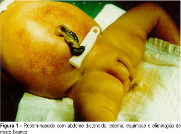
Introduction: meconium peritonitis as result of fetal intestinal perforation has a low incidence (1:30,000 deliveries) and high mortality (50% or more). Prenatal ultrasound findings include fetal ascites and intra-abdominal calcifications. Evidence suggests that prenatal diagnosis can improve postnatal prognosis. Case Report: R.C.M.S., 22 years, II pregnancy O para, presented ultrasound (12/02/98) with diagnosis of fetal ascites. Investigation for hydrops fetalis was performed and immune and nonimmune causes were excluded. Severe fetal ascites persisted on subsequent ultrasound examinations, without calcifications. Vaginal delivery occurred at 36 weeks (01/02/99), with polyhydramnios. Female neonate weighing 2,670 g, with signs of respiratory distress, abdominal distension and petechiae. Abdominal distension worsened progressively, with palpation of a petrous tumor in the right upper quadrant and elimination of white mucus at rectal examination. Radiological findings (01/04/99) were disseminated abdominal calcifications, intestinal dilatation and absence of gas at rectal ampulla. Exploratory laparotomy was indicated with diagnosis of meconium peritonitis. A giant meconium cyst and ileal atresia were observed and lysis of adhesions and ileostomy were performed. Initial postoperative evolution was satisfactory but was subsequently complicated by sepsis and neonatal death occurred (01/09/99). Conclusion: meconium peritonitis should be remembered at differential diagnosis of fetal ascites. In the present case, surgical indication could be anticipated if prenatal diagnosis were established, with improvement of neonatal evolution.
Search
Search in:


Comments