Revista Brasileira de Ginecologia e Obstetrícia. 2003;25(4):249-254

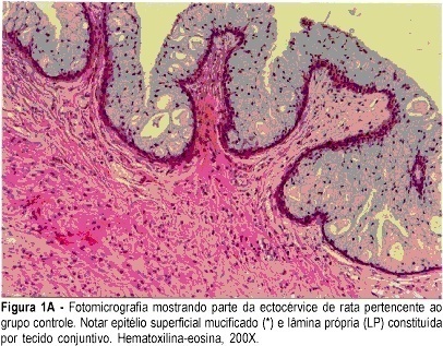
PURPOSE: to assess the morphological and morphometric alterations in the uterine cervix of pregnant albino rats determined by local hyaluronidase administration. METHODS: ten rats with a positive pregnancy test were randomly distributed into two equal groups. The control group consisted of rats that received a single dose of 1 mL distilled water in the uterine cervix, on gestational day 18, under anesthesia. The experimental group consisted of rats that received 0.02 mL hyaluronidase, diluted in 0.98 ml distilled water (total = 1 mL), in the same conditions as those of the control group. On day 20, the rats were anesthetized and submitted to dissection. The uterine cervix was prepared for morphological and morphometric study at light microscopy (hematoxylin and eosin, and Masson trichrome). RESULTS: in the experimental group, greater thinning of the superficial mucified epithelium was observed, with lamina propria rich in blood vessels and eosinophils. Diversely, the control group showed a large concentration of collagen fibers. The histometric analysis in the experimental group was characterized by a smaller number of collagen fibers (mean = 248 versus 552 of control; SD = 49.7 versus 31.1 of control). The parametric method (Student’s t test) showed a significant difference between groups (p<0.0001). CONCLUSION: the local use of hyaluronidase in the cervix of pregnant rats determined predominance of loose connective tissue and a smaller concentration of collagen fibers.
Search
Search in:


Comments