Revista Brasileira de Ginecologia e Obstetrícia. 2003;25(2):115-121

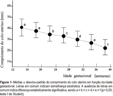
PURPOSE: to establish a normality curve of cervical length during pregnancy measured by transvaginal ultrasonography. METHODS: we conducted a prospective, longitudinal study on 82 healthy pregnant women who were followed up from the beginning of pregnancy to delivery at four-week intervals, of whom 49 concluded the study. Patients were divided according to parity into nulliparous women and women with one or more previous deliveries. Cervical length was measured in a sagittal view by transvaginal ultrasonography, as the linear distance between internal and external cervical os. RESULTS: no significant difference was observed in mean cervical length or the 5th, 25, 50th, 75th, or 95th percentile according to gestational age between groups (p>0.05). Between the 20thand 24th gestacional week, the 5th, 50th and 95th percentiles of cervical length were 28, 35 and 47.2 mm, respectively. Cervical length decreased progressively during normal pregnancy, with a significant shortening observed after 20 weeks of gestation and being more marked after 28 weeks (p<0.05). CONCLUSION: the pattern of cervical length behavior does not seem to differ between nulliparous women and women with one or more previous deliveries. The numerical values of the normality curve of cervical length according to gestational age reflect the variability in the peculiar characteristics of the studied sample, thus emphasizing the value of the parameters established for different populations.
Search
Search in:


Comments