Revista Brasileira de Ginecologia e Obstetrícia. 2005;27(3):137-142

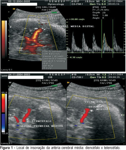
PURPOSE: to evaluate if there is any difference between Doppler indexes in the middle cerebral artery in two different sites of insonation in healthy patients and in patients with diseases. METHODS: a random prospective survey, in the period from June 2003 to March 2004 that analyzed the Doppler indexes of 100 patients: patient group (n = 50) included patients admitted to Clemente Farias University Hospital, which is part of UNIMONTES-MG, havinfg as inclusion criteria: to be in the 28th to 34th gestational week, diagnosis of chronic arterial hypertension, pre-eclampsia, intrauterine growth restriction. As control group, 50 healthy pregnant patients between the 28th and the 34th week, originary from SEMESP’s clinic. The Doppler variables were the resistance index (RI), the pulsatility index (PI) and the relation systole/diastole (SD). All three Doppler indexes were assessed at two different sites of the cerebral artery: the first measurement in the diencephalons region, soon after the beginning of the middle cerebral artery and the second on a distal location in the telencephalon. The median Doppler indexes in the patient group in the first and second measurements were 1.55 and 1.69 for the PI, 0.77 and 0.79 for RI and 4.29 and 4.86 for SD, respectively. In the control group, the values were 1.73 and 1.86 for the PI; 0.83 and 0.79 for RI and 5.83 and 5.46 for SD. There were no differences between sites with a p value of 0.38, 0.29 and 0.39 for PI, RI and SD, respectively. In 15t fetuses with centralization (brain sparing effects), in the diencephalon the median indexes were 1.02 for PI, 0.63 for RI and 2.68 for SD. In the epencephalon the median indexes were 0.95 for IP, 0.62 for RI and 2.44 for SD. There were no differences between sites, with a p value of 0.53 for PI; 0.56 for IR and 0.31 for SD. The Doppler index site of assessment in the middle cerebral arteries does not interfere with the results.
Search
Search in:


Comments