Revista Brasileira de Ginecologia e Obstetrícia. 2014;36(8):359-366
To evaluate the effects of ovariectomy and the hyperprolactinemia procedure in the tibial epiphyseal growth plate of female mice.
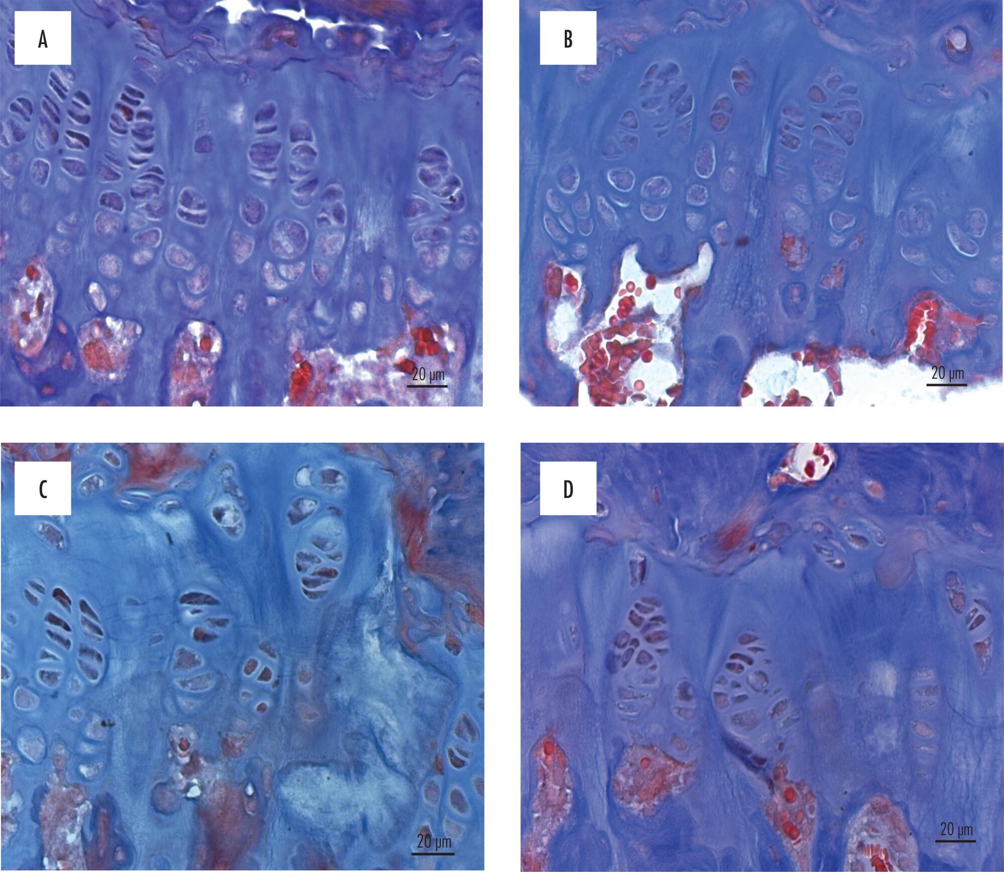
In this study, the epiphyseal growth plate of ovariectomized (OVX) and/or rendered hyperprolactinemic female mice by 50 days of treatment with 200 μg metoclopramide (M) was evaluated morphologically, morphometrically and immuno-histochemically. Forty female and adult mice were divided into four groups according to treatment: V group – animals treated with saline solution; H group – hyperprolactinemic animals; Ovx/V group – ovariectomized animals and treated with saline solution; Ovx/H group – hyperprolactinemic and ovariectomized animals. After the treatment period, the animals were sacrificed, tibia was removed and fixed in 10% buffered formalin and decalcified in 10% formic acid. The material was immersed in paraffin and subjected to histological processing in paraffin. The sections were stained with Masson’s trichrome and immunohistochemistry was carried out for the pro-apoptotic protein BCL-2. The images for the morphological and morphometric study were analyzed with the imaging program AxioVision 4.8 (Carl-Zeiss(r), Germany).
The combination of hyperprolactinemia and the ovariectomy procedure decreased the number of resting chondrocytes 1.5-fold, the number of proliferative chondrocytes 1.8-fold; the percentage of resting cartilage 2.4-fold and the percentage of trabecular bone 2.1-fold, compared with respective control animals.
The procedure of ovariectomy combined with the metoclopramide-induced hyperprolactinemia in female mice has showed marked bone degeneration due to significant decrease of cell proliferation in the epiphyseal growth plate and bone formation.
Search
Search in:
Search
Search in:


Comments