Summary
Revista Brasileira de Ginecologia e Obstetrícia. 2006;28(10):590-595
DOI 10.1590/S0100-72032006001000004
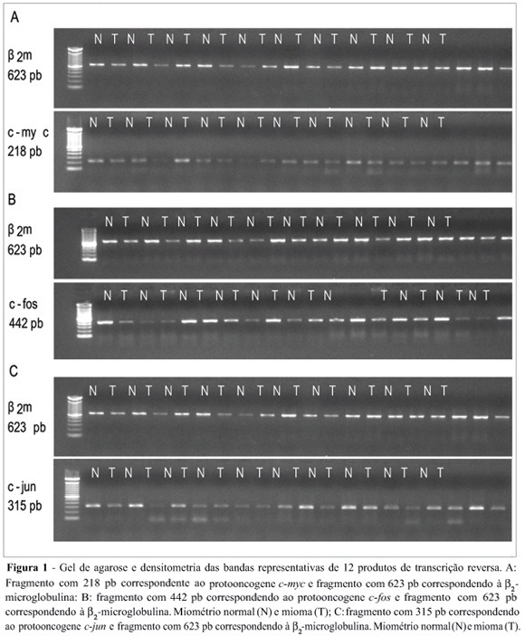
Uterine myomas are common benign tumors of the female genital tract. The expression of growth factor signal transduction cascade components including the protooncogenes c-myc, c-fos, and c-jun seem to be involved in the development of myomas. PURPOSE: To compare the gene (mRNA) and protein expression of the protooncogenes c-fos, c-myc, and c-jun in human normal myometrium and leiomyoma. METHOD: A case-control study was performed. Samples were collected from 12 patients submitted to hysterectomy at the Hospital de Clínicas at Porto Alegre. The expression of the specific mRNA for c-myc, c-fos, c-jun, and beta-microglobulin was assessed through the RT-PCR technique, using specific primers to each gene. The protein expression of these protooncogenes was evaluated through the Western blot technique with specific antibodies. RESULTS: No statistically significant difference was observed in the gene expression for these protooncogenes between normal myometrium and leiomyoma (c-myc: 0,87 ± 0,08 vs 0,87 ± 0,08, p = 0,952; c-fos: 1,10 ± 0,17 vs 1,01 ± 0,11, p = 0,21; c-jun: 1,03 ± 0,12 vs 0,96 ± 0,09, p = 0,168, respectively). No statiscally significant difference was observed for the protein expression of these protooncogenes between normal myometrium and leiomyoma (c-myc: 1,36 ± 0,48 vs 1,53 ± 0,29, p = 0,569; c-fos: 8,85 ± 5,5 vs 6,56 ± 4,22, p = 0,434; e c-jun: 6,47 ± 3,04 vs 5,42 ± 2,03, p = 0,266, respectively). CONCLUSION: No difference was observed in the gene expression (transcription) nor in the protein expression (translation) of the protooncogenes c-myc, c-fos, and c-jun between leiomyoma and myometrium.
Summary
Revista Brasileira de Ginecologia e Obstetrícia. 2006;28(10):590-595
DOI 10.1590/S0100-72032006001000004
Uterine myomas are common benign tumors of the female genital tract. The expression of growth factor signal transduction cascade components including the protooncogenes c-myc, c-fos, and c-jun seem to be involved in the development of myomas. PURPOSE: To compare the gene (mRNA) and protein expression of the protooncogenes c-fos, c-myc, and c-jun in human normal myometrium and leiomyoma. METHOD: A case-control study was performed. Samples were collected from 12 patients submitted to hysterectomy at the Hospital de Clínicas at Porto Alegre. The expression of the specific mRNA for c-myc, c-fos, c-jun, and beta-microglobulin was assessed through the RT-PCR technique, using specific primers to each gene. The protein expression of these protooncogenes was evaluated through the Western blot technique with specific antibodies. RESULTS: No statistically significant difference was observed in the gene expression for these protooncogenes between normal myometrium and leiomyoma (c-myc: 0,87 ± 0,08 vs 0,87 ± 0,08, p = 0,952; c-fos: 1,10 ± 0,17 vs 1,01 ± 0,11, p = 0,21; c-jun: 1,03 ± 0,12 vs 0,96 ± 0,09, p = 0,168, respectively). No statiscally significant difference was observed for the protein expression of these protooncogenes between normal myometrium and leiomyoma (c-myc: 1,36 ± 0,48 vs 1,53 ± 0,29, p = 0,569; c-fos: 8,85 ± 5,5 vs 6,56 ± 4,22, p = 0,434; e c-jun: 6,47 ± 3,04 vs 5,42 ± 2,03, p = 0,266, respectively). CONCLUSION: No difference was observed in the gene expression (transcription) nor in the protein expression (translation) of the protooncogenes c-myc, c-fos, and c-jun between leiomyoma and myometrium.
Summary
Revista Brasileira de Ginecologia e Obstetrícia. 2006;28(10):581-589
DOI 10.1590/S0100-72032006001000003
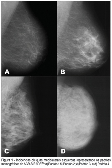
PURPOSE: To assess the presence of estrogen receptor gene polymorphisms HaeIII and MspI as well as clinical factors, and their possible associations with high mammographic density in post-menopausal women. METHODS: One hundred and fifteen post-menopausal women, not in use of hormonal therapy and without clinical or mammographic lesions were evaluated. Three independent observers have determined the mammographic density pattern based on the ACR-BIRADS® 2003 (two subjective and one objective evaluations - Adobe Photoshop 7.0 software). Oral swabs (Cytobrush) were obtained to extract DNA and the polymerase chain reaction - restriction fragment length polymorphism) was performed to assess the presence of polymorphisms in intron 1 and exon 1 from estrogen receptor gene (HaeIII and MspI). RESULTS: The HaeIII polymorphism was found in 43 (37.4%) of the 115 women, while MspI was found in 96 (83.5%) of them. There was a good agreement among determinations of the three observers with regard to mammographic density. Thirty-four (29.6%) women had dense breasts and eighty-one (70.4%) had non-dense breasts. CONCLUSION: The estrogen receptor gene polymorphism Haelll showed no association with mammographic density (Fisher = 0.712), while the association between estrogen receptor gene polymorphism Mspl and mammographic density was near significance (Fisher = 0.098). The associations among age, parity and body mass index revealed statistical significance.
Summary
Revista Brasileira de Ginecologia e Obstetrícia. 2006;28(10):581-589
DOI 10.1590/S0100-72032006001000003
PURPOSE: To assess the presence of estrogen receptor gene polymorphisms HaeIII and MspI as well as clinical factors, and their possible associations with high mammographic density in post-menopausal women. METHODS: One hundred and fifteen post-menopausal women, not in use of hormonal therapy and without clinical or mammographic lesions were evaluated. Three independent observers have determined the mammographic density pattern based on the ACR-BIRADS® 2003 (two subjective and one objective evaluations - Adobe Photoshop 7.0 software). Oral swabs (Cytobrush) were obtained to extract DNA and the polymerase chain reaction - restriction fragment length polymorphism) was performed to assess the presence of polymorphisms in intron 1 and exon 1 from estrogen receptor gene (HaeIII and MspI). RESULTS: The HaeIII polymorphism was found in 43 (37.4%) of the 115 women, while MspI was found in 96 (83.5%) of them. There was a good agreement among determinations of the three observers with regard to mammographic density. Thirty-four (29.6%) women had dense breasts and eighty-one (70.4%) had non-dense breasts. CONCLUSION: The estrogen receptor gene polymorphism Haelll showed no association with mammographic density (Fisher = 0.712), while the association between estrogen receptor gene polymorphism Mspl and mammographic density was near significance (Fisher = 0.098). The associations among age, parity and body mass index revealed statistical significance.
Summary
Revista Brasileira de Ginecologia e Obstetrícia. 2006;28(10):575-580
DOI 10.1590/S0100-72032006001000002
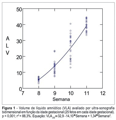
PURPOSE: To determine the values of amniotic fluid in normal fetuses during the first trimester of pregnancy by three- and bi-dimensional ultrasonography. METHODS: In a prospective longitudinal study, 25 normal fetuses were evaluated from the 8th to the 11th week of gestation. Amniotic fluid volume was measured by endovaginal ultrasonography with the three- and two-dimensional modes. The two-dimensional study consisted of volumetric determination by mathematical calculation based on an ellipsoidal shape (constant 0.52) to obtain the amniotic sac and embryo volumes. In the three-dimensional study, the amniotic fluid volume was determined by the VOCAL technique using 6, 9, 15, and 30 degrees of rotation. The amniotic fluid volume obtained by 6-degree rotations was considered to be the final result. In both modes, amniotic fluid volume was obtained by subtracting the volume of the embryo from the volume of the amniotic sac. Data were analyzed statistically for variance (ANOVA), correlation and regression analysis. The level of significance was set at p < 0.05. RESULTS: The amniotic fluid volume as measured by two-dimensional ultrasonography increased from 5.45 to 39.52 cm³ in the range from the 8th to the 11th week (ANOVA - p < 0.05). There was a correlation between gestational age and amniotic fluid volume (p < 0.001, r² = 88.3%). In the three-dimensional study, the amniotic fluid volume increased from 5.7 to 42.9 cm³ in the range from the 8th to the 11th week (ANOVA - p < 0.05), and again a correlation between gestational age and amniotic fluid volume (p < 0.001, r² = 98.1%) was observed. CONCLUSION: an increase in amniotic fluid volume occurs during the first trimester of pregnancy, as determined by the two- and three-dimensional modes. In addition, we have demonstrated that the higher the gestational age, the larger the amniotic fluid volume.
Summary
Revista Brasileira de Ginecologia e Obstetrícia. 2006;28(10):575-580
DOI 10.1590/S0100-72032006001000002
PURPOSE: To determine the values of amniotic fluid in normal fetuses during the first trimester of pregnancy by three- and bi-dimensional ultrasonography. METHODS: In a prospective longitudinal study, 25 normal fetuses were evaluated from the 8th to the 11th week of gestation. Amniotic fluid volume was measured by endovaginal ultrasonography with the three- and two-dimensional modes. The two-dimensional study consisted of volumetric determination by mathematical calculation based on an ellipsoidal shape (constant 0.52) to obtain the amniotic sac and embryo volumes. In the three-dimensional study, the amniotic fluid volume was determined by the VOCAL technique using 6, 9, 15, and 30 degrees of rotation. The amniotic fluid volume obtained by 6-degree rotations was considered to be the final result. In both modes, amniotic fluid volume was obtained by subtracting the volume of the embryo from the volume of the amniotic sac. Data were analyzed statistically for variance (ANOVA), correlation and regression analysis. The level of significance was set at p < 0.05. RESULTS: The amniotic fluid volume as measured by two-dimensional ultrasonography increased from 5.45 to 39.52 cm³ in the range from the 8th to the 11th week (ANOVA - p < 0.05). There was a correlation between gestational age and amniotic fluid volume (p < 0.001, r² = 88.3%). In the three-dimensional study, the amniotic fluid volume increased from 5.7 to 42.9 cm³ in the range from the 8th to the 11th week (ANOVA - p < 0.05), and again a correlation between gestational age and amniotic fluid volume (p < 0.001, r² = 98.1%) was observed. CONCLUSION: an increase in amniotic fluid volume occurs during the first trimester of pregnancy, as determined by the two- and three-dimensional modes. In addition, we have demonstrated that the higher the gestational age, the larger the amniotic fluid volume.
Summary
Revista Brasileira de Ginecologia e Obstetrícia. 2006;28(9):545-550
DOI 10.1590/S0100-72032006000900007
PURPOSE: to evaluate muscular strength of the pelvic floor and the periurethral vessels of postmenopausal women before and after six months of soybean extract treatment. METHODS: the study was conducted on 30 postmenopausal women before and after six consecutive months of soyabean extract (100 mg/day) administration. Urinary loss and muscular strength of the pelvic floor were investigated through digital perineometer and functional evaluation. Digital color Doppler in the periurethral region was used to count the number of vessels. For statistical analysis, the paired Student t test was applied to compare the results before and after the treatment. RESULTS: twenty women reported urinary incontinence before the treatment period. The amelioration of this symptom was observed in 15 (75%) women after the treatment. Vaginal pressure (muscular strength of the pelvic floor) was 12.95±1.73 and 15.86±1.86 Sauers, before and after the treatment, respectively (p<0.001). Twenty-two women (73.3%) presented an increase in the pressure at the end of this study. In relation to the function evaluation, 18 (60%) had improvement in muscular strength and 12 women did not present any change. On ultrasonography (Doppler), the number of vessels was 2.20±0.15 blood vessels/field in the beginning of this study and 3.46±0.25 blood vessels/field at the end of the treatment (p<0.001). An increase in the number of periurethral vessels was detected in 21 women (70%). CONCLUSION: it is important to emphasize that these are preliminary results. A double blind randomized and placebo-controlled clinical trial with a high number of participants is necessary. However, the treatment with concentrated soybean extract (100 mg per day) for six consecutive months may determine an improvement in pelvic floor muscular strength and an increase in the number of periurethral vessels in postmenopausal women.
Summary
Revista Brasileira de Ginecologia e Obstetrícia. 2006;28(9):536-544
DOI 10.1590/S0100-72032006000900006
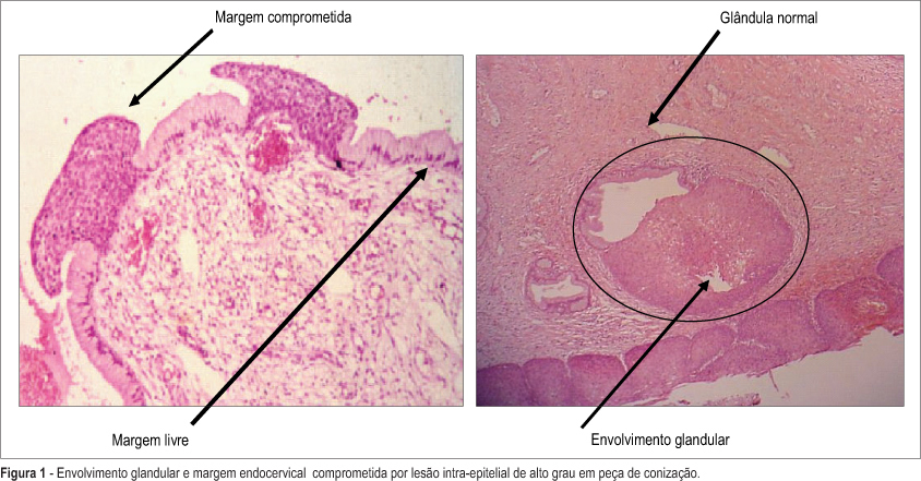
PURPOSE: to evaluate risk factors associated with cervical intraepithelial lesion recurrence after LEEP conization. METHODS: nested case-control study in a cohort of 201 patients with cervical intraepithelial lesion, that were submitted to LEEP conization. Average follow-up of these patients was 2 years. Ninety-four HIV-infected women and 107 non-infected were enrolled. Cervical conization was achieved by the Loop Electrosurgical Excision Procedure (LEEP). Evaluated surgical biopsy histopathological characteristics were lesion grade, lesion borders and glandular involvement. After surgery all patients were submitted to a colposcopy and cytological evaluation every six months. Recurrent lesions were defined it confirmed by biopsy after surgery. Cases were patients with and controls patients without recurrence. chi2 test and multivariable analysis by logistic regression were used for group comparisons. Kaplan Meier's method was performed for the survival analyses (log-rank test). RESULTS: 40 patients had lesion recurrence. Initially, significant variables were: partner number, HIV-infection, lesion borders and glandular involvement. The most frequent recurrence occurred when there were simultaneous association between positive margins and glandular involvement as indicator for recurrence risk. After logistic regression analysis the main factors associated with lesion recurrence were: glandular involvement (OR-9.1; 95% CI:13.0- 27.5); HIV-infection (OR-4.6; 95% IC:1.1-6.3); compromised margins (OR-2.6; 95% IC:1.9-11.2). CONCLUSIONS: risk factors associated with cervical intraepitelial lesion recurrence were HIV-infection, glandular involvement and compromised margins.
Summary
Revista Brasileira de Ginecologia e Obstetrícia. 2006;28(9):536-544
DOI 10.1590/S0100-72032006000900006
PURPOSE: to evaluate risk factors associated with cervical intraepithelial lesion recurrence after LEEP conization. METHODS: nested case-control study in a cohort of 201 patients with cervical intraepithelial lesion, that were submitted to LEEP conization. Average follow-up of these patients was 2 years. Ninety-four HIV-infected women and 107 non-infected were enrolled. Cervical conization was achieved by the Loop Electrosurgical Excision Procedure (LEEP). Evaluated surgical biopsy histopathological characteristics were lesion grade, lesion borders and glandular involvement. After surgery all patients were submitted to a colposcopy and cytological evaluation every six months. Recurrent lesions were defined it confirmed by biopsy after surgery. Cases were patients with and controls patients without recurrence. chi2 test and multivariable analysis by logistic regression were used for group comparisons. Kaplan Meier's method was performed for the survival analyses (log-rank test). RESULTS: 40 patients had lesion recurrence. Initially, significant variables were: partner number, HIV-infection, lesion borders and glandular involvement. The most frequent recurrence occurred when there were simultaneous association between positive margins and glandular involvement as indicator for recurrence risk. After logistic regression analysis the main factors associated with lesion recurrence were: glandular involvement (OR-9.1; 95% CI:13.0- 27.5); HIV-infection (OR-4.6; 95% IC:1.1-6.3); compromised margins (OR-2.6; 95% IC:1.9-11.2). CONCLUSIONS: risk factors associated with cervical intraepitelial lesion recurrence were HIV-infection, glandular involvement and compromised margins.
Summary
Revista Brasileira de Ginecologia e Obstetrícia. 2006;28(9):530-535
DOI 10.1590/S0100-72032006000900005
PURPOSE: to compare the incidence of preterm deliveries, and of low birth weight newborns, among primiparous adolescents, from two age groups. METHODS: this is a comparative, cross-sectional clinical study composed of 522 primiparous adolescents whose deliveries occurred at the gestational age of 25 to 42 weeks. The adolescents were divided into 2 groups according to their age; Gprec: from 10 to 15 complete years old (n=104); Gtard: from 16 to 19 complete years old (n=418). The research data were obtained by an individualized, confidential and ethical interview, soon after delivery; and by a written questionnaire with questions about the gestational age in complete weeks, and about the newborns birth weight. The gestational age was calculated at the delivery day, according to the date of the last trustworthy menstrual period, being also confirmed by the earliest pregnancy scanning or by Capurro's index, when there were any doubts about the previously described parameters. All newborns with gestational age under 37 weeks at birth were considered preterm babies. The newborn weight was taken by neonatologists immediately after delivery; all newborns with less than 2,500g were considered to be low weight babies. Thus, we compared prematurity rate and low birth weight among newborns from primiparous puerperal adolescents. The chi² test was used for the statistic analysis and the partition chi² test for the found differences. As the significancy level was 0.05 (alpha =5%), lower levels than that were considered significant. RESULTS: the prematurity rate was not significantly different between the two groups (5.8 and 2.6%). The incidence of low birth weight in Gprec (13.5%) was significantly higher than in Gtard (3.1%). CONCLUSIONS: the group with primiparous adolescents under 15 years old showed a significantly higher risk of low birth weight newborns. However, a statistically significant incidence of prematurity between the groups studied was not verified.
Summary
Revista Brasileira de Ginecologia e Obstetrícia. 2006;28(9):523-529
DOI 10.1590/S0100-72032006000900004
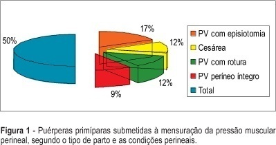
PURPOSE: to determine the values of perineal muscular force (PMF) in the lying and seated positions and to identify the values of PMF between first pregnancy, according to type and the characteristics of the vaginal delivery and cesarean section. METHODS: study of the transversal type, performed in a maternity of Brazilian Public the Health System (SUS) in the city of São Paulo. The sample consisted of 95 primiparae at term. Evaluation occurred between the 40th and 45 th, day with an interview, physical examination and measurement of PMF using a perineometer of the Kegel type. The measurement was carried out in the lying and seated positions, muscular status (at rest and in maximum contraction), and the average of three measures for each position and muscular state were considered. RESULTS: 76.8% (73) of the women had vaginal delivery and 23.2% (22) cesarean section. After vaginal delivery, intact perineum in 18.9%, (18), perineal rupture in 24.2% (23), and episiotomy in 33.7% (32) were observed. Obtained values of the PMF were: lying position muscular rest 18. 9 mmHg, lying position maximum contraction: 30,7 mmHg, seated position muscular rest: 34.5 mmHg, seated positions maximum muscular contraction: 46.5 mmHg. CONCLUSION: there was association between the type and the characteristics of the delivery and PMF.
Summary
Revista Brasileira de Ginecologia e Obstetrícia. 2006;28(9):523-529
DOI 10.1590/S0100-72032006000900004
PURPOSE: to determine the values of perineal muscular force (PMF) in the lying and seated positions and to identify the values of PMF between first pregnancy, according to type and the characteristics of the vaginal delivery and cesarean section. METHODS: study of the transversal type, performed in a maternity of Brazilian Public the Health System (SUS) in the city of São Paulo. The sample consisted of 95 primiparae at term. Evaluation occurred between the 40th and 45 th, day with an interview, physical examination and measurement of PMF using a perineometer of the Kegel type. The measurement was carried out in the lying and seated positions, muscular status (at rest and in maximum contraction), and the average of three measures for each position and muscular state were considered. RESULTS: 76.8% (73) of the women had vaginal delivery and 23.2% (22) cesarean section. After vaginal delivery, intact perineum in 18.9%, (18), perineal rupture in 24.2% (23), and episiotomy in 33.7% (32) were observed. Obtained values of the PMF were: lying position muscular rest 18. 9 mmHg, lying position maximum contraction: 30,7 mmHg, seated position muscular rest: 34.5 mmHg, seated positions maximum muscular contraction: 46.5 mmHg. CONCLUSION: there was association between the type and the characteristics of the delivery and PMF.
Summary
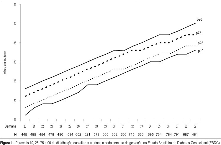
Revista Brasileira de Ginecologia e Obstetrícia. 2006;28(9):513-522
DOI 10.1590/S0100-72032006000900003
PURPOSE: to describe, in participants of the Brazilian Study of Gestational Diabetes (EBDG), the percentile distribution of uterine height by gestational age and to validate the use of percentiles of the chart derived by the "Centro Latino-Americano de Perinatologia" (CLAP), used as reference in predicting abnormal fetal growth. METHODS: the EBDG is a cohort study of 5564 pregnant women older than 19 years, followed through and after delivery. Interviews and standardized anthropometry were performed at baseline between 20-28 weeks. Medical records covering prenatal and delivery periods were then reviewed following a standardized approach. Analyses pertain to 3539 women with gestational age confirmed by ultrasound. Diagnostic properties of the 10th and the 90th percentiles of both charts (EBDG and CLAP) as predictors of abnormal neonatal weight were determined. RESULTS: uterine height was higher in EBDG than in the CLAP chart at every gestational week, being 1-4 and 2-6 cm greater, at the 10th and 90th percentiles respectively. The CLAP 10th percentile classified as small the uterine heights of only 0.3 to 1.7% of Brazilian women, while the 90th percentile classified as large the uterine heights of 42 to 57% of the sample. The sensitivity of CLAP percentile 10 in the prediction of small for gestational age varied from 0.8 to 6% and the specificity of CLAP percentile 90 in the prediction of large for gestational age, from 46 to 61%. CONCLUSIONS: the CLAP uterine height reference chart does not reflect the current uterine growth pattern of pregnant Brazilians, limiting its clinical applicability in the detection of abnormal fetal growth, especially intrauterine growth restriction.
Summary
Revista Brasileira de Ginecologia e Obstetrícia. 2006;28(9):513-522
DOI 10.1590/S0100-72032006000900003
PURPOSE: to describe, in participants of the Brazilian Study of Gestational Diabetes (EBDG), the percentile distribution of uterine height by gestational age and to validate the use of percentiles of the chart derived by the "Centro Latino-Americano de Perinatologia" (CLAP), used as reference in predicting abnormal fetal growth. METHODS: the EBDG is a cohort study of 5564 pregnant women older than 19 years, followed through and after delivery. Interviews and standardized anthropometry were performed at baseline between 20-28 weeks. Medical records covering prenatal and delivery periods were then reviewed following a standardized approach. Analyses pertain to 3539 women with gestational age confirmed by ultrasound. Diagnostic properties of the 10th and the 90th percentiles of both charts (EBDG and CLAP) as predictors of abnormal neonatal weight were determined. RESULTS: uterine height was higher in EBDG than in the CLAP chart at every gestational week, being 1-4 and 2-6 cm greater, at the 10th and 90th percentiles respectively. The CLAP 10th percentile classified as small the uterine heights of only 0.3 to 1.7% of Brazilian women, while the 90th percentile classified as large the uterine heights of 42 to 57% of the sample. The sensitivity of CLAP percentile 10 in the prediction of small for gestational age varied from 0.8 to 6% and the specificity of CLAP percentile 90 in the prediction of large for gestational age, from 46 to 61%. CONCLUSIONS: the CLAP uterine height reference chart does not reflect the current uterine growth pattern of pregnant Brazilians, limiting its clinical applicability in the detection of abnormal fetal growth, especially intrauterine growth restriction.