Summary
Revista Brasileira de Ginecologia e Obstetrícia. 2007;29(7):340-345
DOI 10.1590/S0100-72032007000700003
PURPOSE: to evaluate the efficacy of in vitro fertilization (IVF) with intracytoplasmic sperm injection (ICSI) in natural cycle (NC). METHODS: retrospective clinical trial that evaluated 70 treatment cycles in 60 couples that were submitted to IVF treatment with ICSI in NC performed in private clinic from 1999 until 2003. It was performed daily ultrasound monitorization or on alternate days, and urinary LH dosage when the follicle reached 16 mm of diameter. It was scheduled egg retrieval when the follicle reached 18 mm of diameter and 36 hours after hCG administration when the LH test was negative. Embryo transfer was performed 48 to 52 hours after ICSI. RESULTS: 70 ICSI cycles in 60 patients were performed and the indications of treatment included: male factor (47.1%), tubal factor (37.1%), associated factors (8.7%), unknown infertility (7.1%). Out of 70 cycles, 18 cycles were cancelled (25.7% of cancellation rate). Out of 52 patients that were submitted to ovarian punction to oocyte retrieval we found mature oocytes in 77% of the cases (40 cycles), in four cases we collected immature oocytes and in eight cases we could not found it. We had 70% of fertilization rate and only one fertilized oocyte did not achieve the cleavage stage. So, the transfers rate per punction and per mature oocyte was 52% and 67.5%, respectively. We had 11.4% of pregnancy rate per cycle, 15.4% per punction and 29.6% per embryo transfer. CONCLUSIONS: FIV/ICSI in NC seem to be a satisfactory option of treatment, with low costs and complications (multiple gestation and Ovarian Hyperstimulation Syndrome), mainly in poor responder patients and in poor populations.
Summary
Revista Brasileira de Ginecologia e Obstetrícia. 2007;29(7):335-339
DOI 10.1590/S0100-72032007000700002
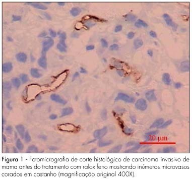
PURPOSE: to evaluate the effect of raloxifene on breast cancer angiogenesis of menopausal women. METHODS: sixteen menopausal women with stage II (>3 cm) estrogen receptor positive operable breast cancer were enrolled in this study. Following confirmation of the diagnosis by incisional biopsy, the patients received 60 mg raloxifene daily for 28 days prior to the definitive surgery. Immunohistochemical study was performed on the sample tumors obtained during the biopsy for the diagnosis and evaluation of the status of estrogen receptor and during the definitive surgery. The anti-CD34 monoclonal antibody was used as a marker for endothelial cells. The vascular unit was considered as any endothelial cell or group of cells of a brownish color, clearly separated from adjacent microvessels, tumor cells or other connective tissue, forming or not lumen. Microvessel count was performed in ten fields of each slide using a 40X objective lens (400X magnification). A microscope coupled to a system of capture and analysis of image was used (Imagelab®). Statistical analysis of data was carried out using the paired Student t-test and significance level was established at p<0.05. RESULTS: mean numbers of anti-CD34 antibody-stained microvessels before and after raloxifene treatment were 44.4±3.5 and 22.6±1.6, respectively. A significant reduction in the number of microvessels following raloxifene therapy was observed (p<0.001). CONCLUSIONS: when administered as primary therapy for menopausal women with breast carcinoma, raloxifene significantly reduced tumoral angiogenesis.
Summary
Revista Brasileira de Ginecologia e Obstetrícia. 2007;29(7):335-339
DOI 10.1590/S0100-72032007000700002
PURPOSE: to evaluate the effect of raloxifene on breast cancer angiogenesis of menopausal women. METHODS: sixteen menopausal women with stage II (>3 cm) estrogen receptor positive operable breast cancer were enrolled in this study. Following confirmation of the diagnosis by incisional biopsy, the patients received 60 mg raloxifene daily for 28 days prior to the definitive surgery. Immunohistochemical study was performed on the sample tumors obtained during the biopsy for the diagnosis and evaluation of the status of estrogen receptor and during the definitive surgery. The anti-CD34 monoclonal antibody was used as a marker for endothelial cells. The vascular unit was considered as any endothelial cell or group of cells of a brownish color, clearly separated from adjacent microvessels, tumor cells or other connective tissue, forming or not lumen. Microvessel count was performed in ten fields of each slide using a 40X objective lens (400X magnification). A microscope coupled to a system of capture and analysis of image was used (Imagelab®). Statistical analysis of data was carried out using the paired Student t-test and significance level was established at p<0.05. RESULTS: mean numbers of anti-CD34 antibody-stained microvessels before and after raloxifene treatment were 44.4±3.5 and 22.6±1.6, respectively. A significant reduction in the number of microvessels following raloxifene therapy was observed (p<0.001). CONCLUSIONS: when administered as primary therapy for menopausal women with breast carcinoma, raloxifene significantly reduced tumoral angiogenesis.
Summary
Revista Brasileira de Ginecologia e Obstetrícia. 2007;29(6):317-323
DOI 10.1590/S0100-72032007000600007
PURPOSE: to evaluate the diagnostic efficacy of core needle biopsy in suspicious solid breast masses (Bi-Rads™ 4 or 5) up to 10 mm. METHODS: this study was conducted on 83 women diagnosed with 90 suspicious solid lumps, all measuring up to 10 mm, identified by mammography (41), sonography (47) or palpation (two), who visited our clinic from September 2003 to January 2006. The lesions were biopsied with automatic device and 14 gauge needle (Bard, Corington, GA), all of them ultrasound guided. According to the pathological report, the biopsies were classified as true-positives, false-positives, true-negatives, and false-negatives. The sensitivity and specificity of ultrasound-guided core biopsy in breast masses up to 1 cm were calculated. RESULTS: the mean patient age was 52.5 years, and the mean diameter of the lesions was 7.3 mm, ranging from 3 to 10 mm. The results yielded by the pathologist showed 40 benign lesions (44.4%), 49 malignant lesions (54.4%) and one case (1.1%) of inconclusive material. This patient was submitted to ultrasound guided core needle biopsy once again, this time with conclusive results. There was agreement between histology and radiology in 89 cases. Sensitivity of ultrasound guided core biopsy for the diagnosis of breast cancer was 98.8 % and specificity 100%. CONCLUSIONS: the results obtained by ultrasound-guided core biopsy in suspicious breast lesions up to 10 mm are comparable to the best results of any kind of biopsy in the literature. Therefore, even solid lesions smaller or equal to 10 mm can be safely evaluated pathologically by core biopsy.
Summary
Revista Brasileira de Ginecologia e Obstetrícia. 2007;29(6):310-316
DOI 10.1590/S0100-72032007000600006
PURPOSE: to evaluate the expression of cyclooxygenase-2 (COX-2) in ductal carcinoma in situ (DCIS), invasive ductal carcinoma (IDC), adjacent normal stroma, and epithelium. The correlation of expression levels with nuclear grade, histological grade, presence or absence of comedonecrosis, tumor size, and patient age was also analyzed. METHODS: forty-seven surgical samples obtained from mastectomy and quadrantectomy with simultaneous DCIS and IDC, stages I and II were included. Anti-COX-2 polyclonal antibodies were used to determine enzyme expression. Samples were classified from zero to three, in accordance with number and intensity of stained cells. RESULTS: COX-2 was positively expressed in IDC, DCIS, and normal epithelium in 86.7, 84.4, and 73.3% of the cases, respectively. Concerning nuclear grade (NG), COX-2 expression was positive in 80% of cases of NG-I; in 81.5 and 78.9% of NG II, and in 88.5 and 96.1% of NG III in DCIS and IDC, respectively. COX-2 expression occurred in 78.9% of DCIS with comedonecrosis and in 89.3% without comedonecrosis. As to histological grade (HG) of IDC, COX-2 was positive in 83.3% of HG-I; 89.9% of HG-II and 80% of HG-III. Concerning tumor diameter, COX-2 was present in 86.1% of IDC cases and in 83.3% of DCIS larger than 2 cm and in 11% of IDC and DCIS tumors ≤2 cm. The age range ≥50 years presented 90% expression for IDC and 86.7% for DCIS, and the expression was 92.5% for both IDC and DCIS in patients <50 years. CONCLUSIONS: our results demonstrated high correlation between COX-2 expression in IDC, DCIS and in the normal epithelium, which is consistent with the hypothesis that COX-2 over expression is an early event in breast carcinogenesis. There was no significant correlation between COX-2 expression in IDC and DCIS and nuclear grade, histological grade, presence of comedonecrosis, age group and tumor size.
Summary
Revista Brasileira de Ginecologia e Obstetrícia. 2007;29(6):303-309
DOI 10.1590/S0100-72032007000600005
PURPOSE: to assess the level of lipid peroxidation (LP) and vitamin E in the follicular fluid and serum of infertile patients, with or without endometriosis, submitted to induction of ovulation for assisted reproduction procedures. METHODS: infertile patients aged 20 to 38 years old were selected prospectively and consecutively and divided into Endometriosis Group (17 patients with pelvic endometriosis) and Control Group (19 patients with previous tubal ligation or with male factor). Blood samples were collected on: D1 (before the beginning of the use of gonadotrophins), D2 (day of human chorionic gonadotrofin application) and D3 (day of oocyte retrieval). On D3, follicular fluid samples free from blood contamination were also collected and stored. LP was assessed for malondialdehyde (MDA) quantification by spectrophotometry, and antioxidant status by measurement of vitamin E by HLPC. RESULTS: on D1, no significant difference in LP was observed between groups. However, vitamin E levels were significantly higher in the Control Group. On D2, LP levels were significantly higher in the Endometriosis Group compared to Control and vitamin E levels continued to be significantly higher in the Control Group. On D3, there was no significant difference in both serum and follicular fluid levels of LP or vitamin E between groups. However, on D3, vitamin E levels were found to be significantly higher in serum than in follicular fluid in both groups, whereas MDA levels were significantly lower in follicular fluid than in serum only in the Control Group. CONCLUSION: before the beginning of the induction of ovulation, a significant decrease in antioxidant status was observed in patients with endometriosis, perhaps because antioxidants are consumed during oxidation reactions. After the induction of ovulation with exogenous gonadotrophins, the group of patients with endometriosis presented not only increased lipid peroxidation compared to Control, but also maintained a lower antioxidant status than the Control Group. However, on the day of oocyte retrieval, both serum LP potential and the levels of vitamin E were found to be similar in both groups.
Summary
Revista Brasileira de Ginecologia e Obstetrícia. 2007;29(6):297-302
DOI 10.1590/S0100-72032007000600004
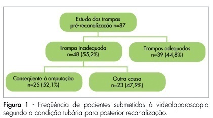
PURPOSE: to identify the main characteristics of the diagnostic and surgical gynecological laparoscopies carried out in patients with reproductive difficulties at a teaching hospital in Recife, from 2000 and 2004. METHODS: a hospital based descriptive case-series study was carried out with 295 patients who had undergone gynecological laparoscopy for either infertility or tube recanalization in the Mother and Child Health Professor Fernando Figueira Institute. Information was obtained from the surgical records of the laparoscopies carried out from January 2000 to December 2004. The inclusion criteria was infertility or pre-recanalization study as a surgical indication. The information was typed twice into a data bank. Tables with central measurements and dispersion tendency were created for the quantitative variables and frequency distribution for the categorical variables. The statistical program, Epi Info 3.3.2., was used to analyze the data. RESULTS: along the study, 462 gynecological laparoscopies were analyzed, 295 (63.8%) of them having as an indication either infertility (41.1%) or the study of possible tube recanalization (18.8%). The patients’ average age in both groups was from 30 to 34 years old. Among the 87 patients with desire of tube recanalization, 55.2% had one or both tubes inadequate for the procedure, and from those, 52.1% was diagnosed with tube amputation (fimbrectomy). In the infertility cases, the most observed findings were adherences (60.6%), tube obstruction (40.9%) and endometriosis (36.1%). Among the procedures carried out, lysis of adherences (34.2%) and biopsies (21%) were the most frequent, followed by endometriosis treatment (10.8%) and salpingostomy (10.8%). CONCLUSION: videolaparoscopy is an important tool in the study and treatment of patients with infertility and before tube recanalization, especially in those hospitals where advanced reproductive techniques are not available.
Summary
Revista Brasileira de Ginecologia e Obstetrícia. 2007;29(6):297-302
DOI 10.1590/S0100-72032007000600004
PURPOSE: to identify the main characteristics of the diagnostic and surgical gynecological laparoscopies carried out in patients with reproductive difficulties at a teaching hospital in Recife, from 2000 and 2004. METHODS: a hospital based descriptive case-series study was carried out with 295 patients who had undergone gynecological laparoscopy for either infertility or tube recanalization in the Mother and Child Health Professor Fernando Figueira Institute. Information was obtained from the surgical records of the laparoscopies carried out from January 2000 to December 2004. The inclusion criteria was infertility or pre-recanalization study as a surgical indication. The information was typed twice into a data bank. Tables with central measurements and dispersion tendency were created for the quantitative variables and frequency distribution for the categorical variables. The statistical program, Epi Info 3.3.2., was used to analyze the data. RESULTS: along the study, 462 gynecological laparoscopies were analyzed, 295 (63.8%) of them having as an indication either infertility (41.1%) or the study of possible tube recanalization (18.8%). The patients’ average age in both groups was from 30 to 34 years old. Among the 87 patients with desire of tube recanalization, 55.2% had one or both tubes inadequate for the procedure, and from those, 52.1% was diagnosed with tube amputation (fimbrectomy). In the infertility cases, the most observed findings were adherences (60.6%), tube obstruction (40.9%) and endometriosis (36.1%). Among the procedures carried out, lysis of adherences (34.2%) and biopsies (21%) were the most frequent, followed by endometriosis treatment (10.8%) and salpingostomy (10.8%). CONCLUSION: videolaparoscopy is an important tool in the study and treatment of patients with infertility and before tube recanalization, especially in those hospitals where advanced reproductive techniques are not available.
Summary
Revista Brasileira de Ginecologia e Obstetrícia. 2007;29(6):291-296
DOI 10.1590/S0100-72032007000600003
PURPOSE: loss of cutaneous sensitivity has been related to lesions of the intercostobrachial nerve (ICBN) during the axillary lymph node dissection for breast cancer treatment. We evaluated pain and cutaneous sensitivity in the ICBN dermatome of patients in which the nerve was preserved during the axillary dissection. METHODS: we carried out a prospective cohort study of 77 patients divided into: NP group (n=34), patients without ICBN preservation, and ICB group (n=43), patients in which the nerve was preserved. Cutaneous sensitivity was evaluated one year after surgery using 1) a modified McGill Pain Questionnaire; 2) clinical examination including brachial perimetry and evaluation of pain and tactile sensitivity; 3) Semmes-Weinstein monofilaments which allow an objective, qualitative, and quantitative evaluation of peripheral nerve lesions. RESULTS: pain was more frequently reported in the NP group (23/33) than in patients from the ICB group (17/42); p=0,012. Painful sensitivity was preserved in the majority of patients from the ICB group (38/42) but in only 11/33 patients from the NP group (p<0,01). There was no significant difference in the number of lymph nodes dissected between the two groups (p=0,06). CONCLUSIONS: patients with ICBN preservation had less pain and more preservation of cutaneous sensitivity, with no decreased number of axillary lymph nodes removed during the axillary dissection.
Summary
Revista Brasileira de Ginecologia e Obstetrícia. 2007;29(6):291-296
DOI 10.1590/S0100-72032007000600003
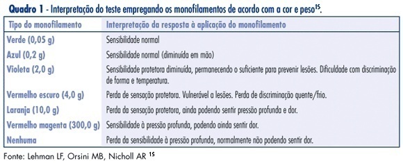
PURPOSE: loss of cutaneous sensitivity has been related to lesions of the intercostobrachial nerve (ICBN) during the axillary lymph node dissection for breast cancer treatment. We evaluated pain and cutaneous sensitivity in the ICBN dermatome of patients in which the nerve was preserved during the axillary dissection. METHODS: we carried out a prospective cohort study of 77 patients divided into: NP group (n=34), patients without ICBN preservation, and ICB group (n=43), patients in which the nerve was preserved. Cutaneous sensitivity was evaluated one year after surgery using 1) a modified McGill Pain Questionnaire; 2) clinical examination including brachial perimetry and evaluation of pain and tactile sensitivity; 3) Semmes-Weinstein monofilaments which allow an objective, qualitative, and quantitative evaluation of peripheral nerve lesions. RESULTS: pain was more frequently reported in the NP group (23/33) than in patients from the ICB group (17/42); p=0,012. Painful sensitivity was preserved in the majority of patients from the ICB group (38/42) but in only 11/33 patients from the NP group (p<0,01). There was no significant difference in the number of lymph nodes dissected between the two groups (p=0,06). CONCLUSIONS: patients with ICBN preservation had less pain and more preservation of cutaneous sensitivity, with no decreased number of axillary lymph nodes removed during the axillary dissection.
Summary
Revista Brasileira de Ginecologia e Obstetrícia. 2007;29(6):285-290
DOI 10.1590/S0100-72032007000600002
PURPOSE: to evaluate the spreading of endometrial cells to the peritoneal cavity during diagnostic hysteroscopy. METHODS: a prospective, descriptive study involving 76 patients divided in two groups: one with 61 patients without malignant endometrial cancer, and the other with 15 patients with endometrial cancer. Two samples of peritoneal fluid were collected, one before (PF-1) and the other immediately after (PF-2) the diagnostic hysteroscopy. Spread to the peritoneal cavity was defined by the presence of endometrial cells in PF-2, with the absence of such cells in PF-1. The 5 mm diameter Storz’s hysteroscopy was used. Distention was obtained by CO2 with electronically controlled flow pressure of 80 mmHg. The PF was fixated in absolute alcohol (ratio1:1). The PF samples were centrifuged and aliquots were smeared and stained using the Papanicolaou method. Analyses were performed by the same observer. RESULTS: during the study, four patients (5.26%) were excluded for presenting endometrial cells in PF-1. In the remaining 72 patients, there was no spread of cells to the peritoneal cavity. In the non-endometrial cancer group, 88.1% (52/59) presented secretory endometrial phase, with correlation of 80% between the hysteroscopy and the biopsy. In the group with endometrial cancer, most of the patients were in stage I (92.3%). There was a 100% correlation between the hysteroscopy/biopsy and histopathology of the surgical sample. CONCLUSIONS: the diagnostic hysteroscopy with CO2 at flow pressure of 80 mmHg did not cause spread of endometrial cells to the peritoneal cavity in both groups, thus suggesting that the diagnostic hysteroscopy is safe for patients at high risk for endometrial cancer.