Summary
Revista Brasileira de Ginecologia e Obstetrícia. 2007;29(8):415-422
DOI 10.1590/S0100-72032007000800006
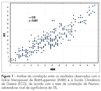
PURPOSE: to evaluate climacteric symptoms and related factors in women living in rural and urban areas of Rio Grande do Norte, Brazil. METHODS: a cross-sectional study involving 261 women in the climacteric was performed. A total of 130 women from Natal and Mossoró (urban group) and 131 from Uruaçu, in São Gonçalo do Amarante (rural group), were studied. Climacteric symptoms were assessed by the Blatt-Kupperman Menopausal Index (BKMI) and Greene Climacteric Scale (GCE). Statistical analysis involved comparison of median between groups and logistic regression analysis. Patients were defined as "very symptomatic" when the climacteric score was >20 for both questionnaires (dependent variable). Independent variables were: age, living area, schooling, obesity and physical activity. RESULTS: the urban group had significantly higher scores than those of the rural group, both for BKMI (median of 26.0 and 17.0, respectively; p<0.0001) and for GCE (median of 27.0 and 16.0, respectively; p<0.0001). For the entire sample, a total of 56.3% (n=147) of the women were classified as "very symptomatic". This prevalence was significantly higher in urban than in rural women (79.2 and 33.6%, respectively; p<0.05). Logistic regression analysis showed that the likelihood of belonging to the group defined as "very symptomatic" was greater for urban women [adjusted odds ratio (OR)=7.1; confidence interval at 95% (95%CI)=3.69-13.66] who were literate (OR=2.19; 95%CI=1.16-4.13). Individuals over the age of 60 years had less chance of having significant symptoms (OR=0.38; 95%CI=0.17-0.87). CONCLUSIONS: the prevalence of significant climacteric symptoms is less in women from a rural environment, showing that sociocultural and environmental factors are strongly related to the appearance of climacteric symptoms in our population.
Summary
Revista Brasileira de Ginecologia e Obstetrícia. 2007;29(8):408-414
DOI 10.1590/S0100-72032007000800005
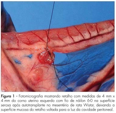
PURPOSE: to analyze the macroscopic and histological changes that occur with the use of sinvastatin in experimental endometriosis in female rats. METHODS: forty Wistar female rats were submitted to the technique of uterine self-transplant in mesenterium. After three weeks, 24 of them developed experimental endometriosis grade III, and were divided in two groups: one group received sinvastatin orally (20 mg/kg/day) and the other (control group) received 0.9% of sodium chloride orally (1 mL/100 g of body weight/day). Both groups received gavage for 14 days, followed by death. The implant volume was calculated [4pi (lenght/2) x (width/2) x (height/2)/3] at the surgical intervention and after the animal’s death. The self-transplants were removed, dyed with hematoxylin-eosin and analyzed by light microscopy. The Mann-Whitney’s test was used in the independent samples and the Wilcoxon’s test for the related samples. The Fisher’s exact test was used for the histological evaluation, with a significance level of 5%. RESULTS: the difference between groups of the initial average volumes of the self-transplants was not significant (p=1.00), but became significant for the final average volumes (p=0.04). There was a significant increase (p=0.01) between the initial and final average volumes in the control group, and a no significant decrease in the sinvastatin group (p=0.95). Histologically, the sinvastatin group (n=9) presented seven cases (77.8%) of moderately preserved and two cases (22.2%) of well preserved epithelial wall, while the control group (n=12) presented seven cases (58.3%) of moderately preserved and five cases (41.7%) of well preserved epithelial wall. CONCLUSIONS: sinvastatin prevented the growth of experimental endometriosis. Studies with sinvastatin for longer periods are promising.
Summary
Revista Brasileira de Ginecologia e Obstetrícia. 2007;29(8):402-407
DOI 10.1590/S0100-72032007000800004
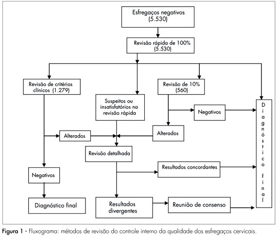
PURPOSE: to evaluate the efficiency of the 100% rapid rescreening in the detection of false-negative results and to verify whether the results vary according to the adequacy of the sample and the woman’s age group. METHODS: to evaluate the efficiency of the rapid rescreening, the 5,530 smears classified as negative by the routine screening, after being submitted to the rapid rescreening of 100%, were compared with the rescreening of the smears on the basis of clinical criteria and 10% random rescreening. For statistical analysis, the variables were evaluated descriptively and the c² test and the Cochran-Armitage test were applied to compare results. RESULTS: of the 141 smears identified as suspicious according to the rapid rescreening method, 84 (59.6%) cases were confirmed in the final diagnosis, of which 36 (25.5%) were classified as atypical squamous cells of undetermined significance, five (3.5%) as atypical squamous cells that cannot exclude high-grade squamous intraepithelial lesion, 34 (24.1%) as low-grade squamous intraepithelial lesion, six (4.3%) as high-grade squamous intraepithelial lesion, and three (2.1%) as atypical glandular cells. Of the 84 suspect smears confirmed in the final diagnosis, 62 (73.8%) smears were classified as adequate and 22 (26.2%) as adequate but with some limitation, but no significant difference was observed with the woman’s age. CONCLUSIONS: the results of this study show that rapid rescreening is an efficient option for internal quality control for the detection of false-negative cervical smear results. In addition, it should be noted that rapid rescreening performed better when the sample was classified as adequate for analysis; however, it did not vary according to the woman’s age group.
Summary
Revista Brasileira de Ginecologia e Obstetrícia. 2007;29(8):387-395
DOI 10.1590/S0100-72032007000800002
PURPOSE: to identify the main maternal risk factors involved in early-onset neonatal sepsis, evaluating the risk associations between bacterial vaginosis and isolated microorganisms found in the maternal urine culture and in the newborn blood culture in the delivery room. METHODS: randomized longitudinal cohort study involving 302 mothers and their newborns. All neonates were followed up for seven days in order to diagnose sepsis. RESULTS: the outcomes were the following: 16 (5.3%) early-onset neonatal sepsis cases (incidence of 53 cases per 1,000 live births). The average number of prenatal appointments with a doctor was 5.2 (SD=1.8). The number of women with prenatal follow-up was 269 (89.1%), but only 117 (43.4%) of them went to six or more medical appointments, 90 (29.8%) had premature rupture of membranes before delivery, but only 22 (7.3%) had it for more than 18 hours. A total of 123 women (40.7%) complained of vaginal discharge, but only 47 (15.6%) of them had bacterial vaginosis, 92 (30.4%) complained of urinary infection, but only 23 (7.6%) of them had bacteriuria, two (0.7%) had fever at home, 122 (40.4%) received intra-partum antibiotic prophylaxis, 40 (13.2%) had premature delivery and 37 (12.3%) had low-birth-weight babies. Gestational age was a significant risk factor (RR=92.9; IC95%:12.6-684.7), as well as the number of prenatal appointments (RR=10,8; IC95%:1,4-80,8), fever (RR=10,0; IC95%:2,3-43,5), low-birth-weight (RR=21,5; IC95%:7,3-63,2) and early neonatal death (RR=89,4; IC95%:11,16-720,6). A significant difference of 5% was found in the comparison of the averages of lower number of prenatal appointments, prematurity and lower birth weight. CONCLUSIONS: the major microorganism isolated in the newborns’ blood culture was the Streptococcus agalactiae. Prematurity, lack of prenatal follow up and low birth weight were the risk factors more associated with early neonatal sepsis.
Summary
Revista Brasileira de Ginecologia e Obstetrícia. 2007;29(7):358-365
DOI 10.1590/S0100-72032007000700006
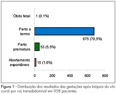
PURPOSE: to evaluate fetal maternal complications after chorionic villus sampling (CVS) for prenatal diagnosis of genetic disorders in pregnant women of Salvador (BA), Brazil. METHODS: case-series study of 958 pregnancies with high risk for chromosomal abnormality submitted to CVS transabdominal between the ninth to the 24th week of gestation, using an ultrasound-guided 18G 3½ spinal needle, from 1990 to 2006. The variables for the analysis of immediate complications were uterine cramps, subchorionic hematoma, accidental amniotic cavity punction, pain in the punction area, amniotic fluid leakage, abdominal discomfort, fetal arrhythmias and vaginal bleeding, and of late complication, abdominal pain, vaginal bleeding, amniotic fluid leakage, infection and spontaneous miscarriage. Premature labor, obstetrical complications (abruption placenta and placenta previa) and newborn malformation were also studied. Qui-square, Student’s "t" or Mann-Whitney tests were used for the statistical analysis; the significance level was 5%. RESULTS: maternal mean age was 36.3±4.9 years old. Immediate complications ware found in 182 (19%) cases (uterine cramp in 14%, subchorionic hematoma in 1.8% and accidental amniotic cavity punction in 1.3%). Late complications were found in 32 (3.3%) cases (vaginal bleeding in 1.6%, abdominal pain in 1.4%, amniotic fluid leakage in 0.3% and spontaneous miscarriage in 1.6% cases). There was no case of abruption placentae, placenta previa or fetal malformation. CONCLUSIONS: CVS is a simple and safe procedure. CVS should be performed in high risk pregnant patients who need prenatal diagnosis of fetal chromosomal abnormalities.
Summary
Revista Brasileira de Ginecologia e Obstetrícia. 2007;29(7):366-369
DOI 10.1590/S0100-72032007000700007
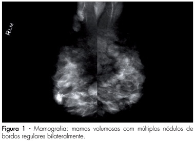
Fibroadenoma is the most frequent benign neoplasia in the female breast and it is considered a mixed tumor, constituted by variable amounts of connective and epithelial tissue. Cyclosporine A seems to be related with the development of mamary fibroadenomas in patients who underwent kidney transplantation in reproductive age. We reported the case in which the patient, in therapeutic use of cyclosporine A, after kidney transplantation, presented several bilateral lumps. The imaging and palpable findings suggested fibroadenoma, confirmed after biopsy.
Summary
Revista Brasileira de Ginecologia e Obstetrícia. 2007;29(7):352-357
DOI 10.1590/S0100-72032007000700005
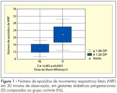
PURPOSE: to analyze the pattern of fetal breathing movements (FBM) in diabetic pregnant women in the third trimester of pregnancy. METHODS: sixteen pregestational diabetic and 16 nondiabetic (control group) pregnant subjects were included fulfilling the following criteria: singleton, between 36-40 weeks of gestation, absence of other maternal diseases and absence of fetal anomalies. The fetal biophysical profile (FBP) was performed to evaluate the following parameters: fetal heart rate, FBM, fetal body movements, fetal tone and amniotic fluid index. The FBM was evaluated for 30 minutes, period when the examination was integrally recorded in VHS video for posterior analysis of the number of FBM episodes, the duration of each episode and the fetal breathing movements index (BMI). The BMI was calculated by the formula: (interval of time with FBM/total time of observation) x 100. At the beginning and in the end of the FBP maternal glucose levels were checked. The results were analyzed by the Mann-Whitney U-test and the Fisher exact test, adopting a level of significance of 5%. RESULTS: the glucose levels demonstrated significantly superior average in the diabetic group (113.3±35.3 g/dL) in relation to the normal group (78.2±14.8 g/dL, p<0.001). The average of the amniotic fluid index was higher in the group of the diabetic cases (15.5±6.4 cm) when compared with controls (10.6±2.0 cm; p=0.01). The average of the number of FBM episodes was superior in the diabetic ones (22.6±4.4) in relation to controls (14.8±2.3; p<0.0001). The average of the BMI in the diabetic patients (54.6±14.8%) was significantly higher than that in the control group (30.5±7.4%, p<0.0001). CONCLUSIONS: the elevated blood glucose levels can be associated with a different pattern in the FBM of diabetic mothers. The use of this parameter of the FBP, in the obstetric practice, must be considered with concern in diabetic pregnancies.
