Summary
Revista Brasileira de Ginecologia e Obstetrícia. 2018;40(10):642-646
Transverse vaginal septum is a rare female genital tract anomaly, and little is described about its surgical treatment. We report the case of a patient who wished to preserve hymenal integrity due to social and cultural beliefs. We performed a vaginoscopic resection of the septum under laparoscopic view, followed by the introduction of a Foley catheter in the vagina, thus preserving the hymen. After 12 months of follow-up, no septal closure was present, and the menstrual flow was effective. Vaginoscopic hysteroscopy is an effectivemethod of vaginal septum resection, even in cases in which hymenal integrity must be preserved due to social and cultural beliefs.
Summary
Revista Brasileira de Ginecologia e Obstetrícia. 2006;28(11):643-651
DOI 10.1590/S0100-72032006001100003
PURPOSE: literature reports show that there are no conclusive data about the association between endometriosis and the concentrations of hormones involved in the control of reproduction. Thus, the present study was undertaken to determine FSH, LH, estradiol (E), progesterone (P), and histamine (Hi) concentrations in serum, peritoneal fluid and follicular fluid of women with and without endometriosis. METHODS: the extent of the disease was staged according to the revised American Fertility Society classification (1997). For the collection of serum and peritoneal fluid, 28 women with endometriosis undergoing diagnostic laparoscopy were selected (18 infertile women with endometriosis I-II and ten infertile women with endometriosis III-IV). For the control group, 21 fertile women undergoing laparoscopy for tubal sterilization were selected. Follicular fluid was obtained from 39 infertile women undergoing in vitro fertilization (21 women with endometriosis and 18 women without endometriosis). RESULTS: FSH and LH levels in serum, peritoneal fluid and follicular fluid did not differ significantly between groups. On the other hand, E and P concentrations in the peritoneal fluid were significantly lower in infertile women with endometriosis (E: 154.2±15.3 for stages I-II and 89.3 ng/mL±9.8 ng/mL for stages III-IV; P: 11.2±1.5 for stages I-II and 7.6 ng/mL±0.8 for stages III-IV) in comparison with control women (E: 289.1 ng/mL±30.1; P: 32.8±4.1 ng/mL) (Kruskal-Wallis/Dunn tests; p<0.05). In serum, estradiol and progesterone concentrations followed the same pattern. In the follicular fluid, E and Hi concentrations were significantly lower in women with endometriosis (E: 97.4±11.1 pg/mL; Hi: 6.6±0.9 ng/mL) in comparison to women without endometriosis (E: 237.5±28.5 pg/mL; Hi: 13.8±1.3 ng/mL) (Student t-test; p<0.05), while progesterone levels revealed no significant difference between groups. CONCLUSIONS: our results indicate ovary dysfunction in women with endometriosis, with reduction on E, P and Hi concentrations, which may contribute to the subfertility often associated with the disease.
Summary
Revista Brasileira de Ginecologia e Obstetrícia. 2017;39(12):645-646
Summary
Revista Brasileira de Ginecologia e Obstetrícia. 2017;39(12):645-646
Summary
Revista Brasileira de Ginecologia e Obstetrícia. 2021;43(9):645-647
Summary
Revista Brasileira de Ginecologia e Obstetrícia. 2021;43(9):645-647
Summary
Revista Brasileira de Ginecologia e Obstetrícia. 2023;45(11):646-653
Currently, uteroplacental vascular disorders are considered one of the main mechanisms of spontaneous preterm delivery (PTD). Low-dose aspirin is used to prevent pre-eclampsia, which has a similar mechanism; hence, the present study aimed to investigate the effect of low-dose aspirin on the prevention of PTD in women with a history of spontaneous PTD.
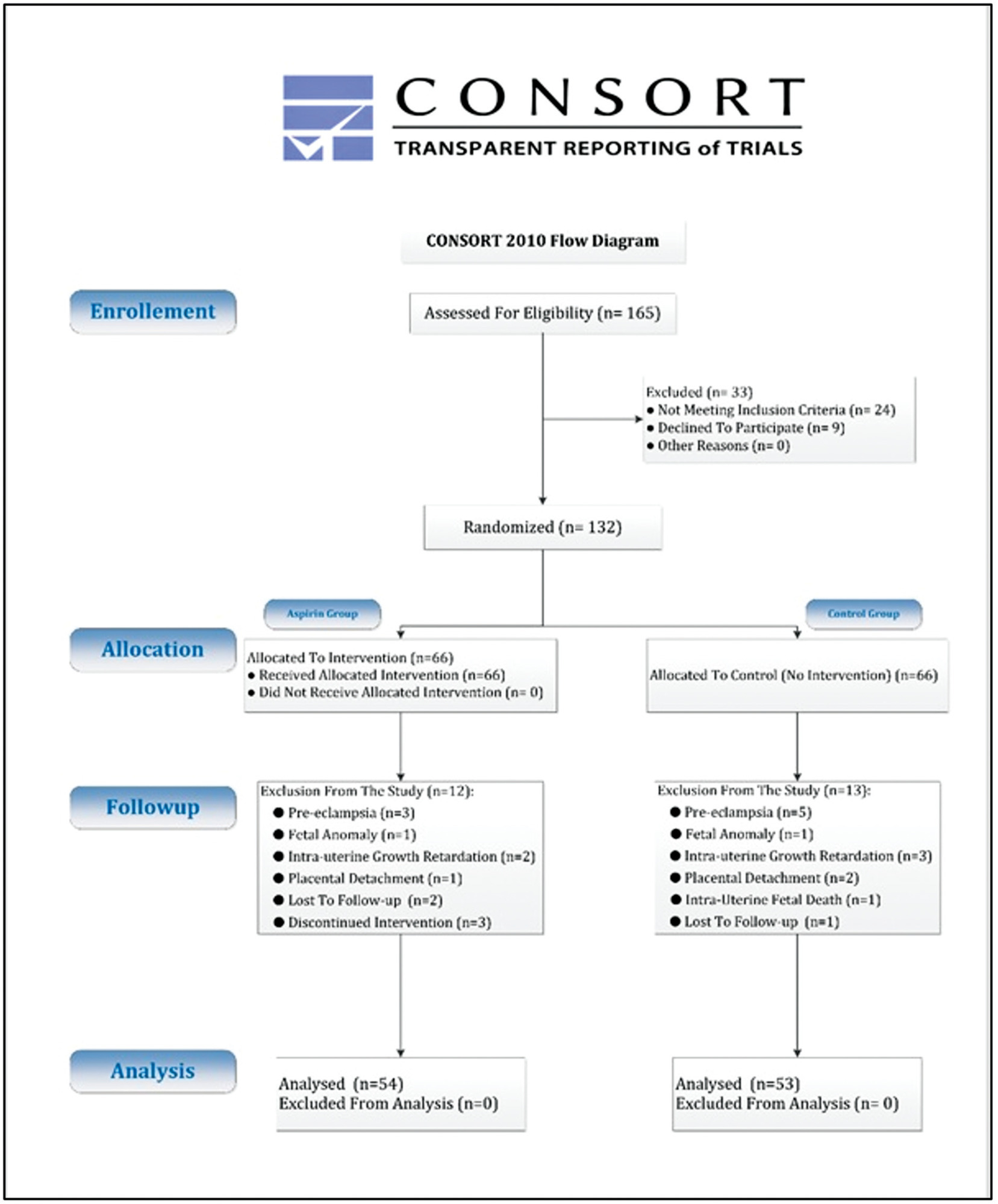
The present pilot randomized clinical trial was conducted on 54 pregnant women in the aspirin group (taking 80 mg daily until the 36th week and classic treatment) and 53 patients in the control group (only receiving classic treatment).
Forty-three patients (40%) presented before 37 weeks due to symptoms of PTL. Preterm delivery (< 37 weeks) occurred in 28 patients (26%), and there was no significant difference between the aspirin and control groups (10 patients [19%] and 18 patients [34%], respectively; p = 0.069). The time of preterm delivery was early (< 34 weeks) in 6 patients (21%), and its cause was spontaneous labor in 23 patients (82%) which was not significantly different between the two groups (p > 0.05). Out of 40 patients with spontaneous labor, 25 patients (63%) had a PTD, which was significantly lower in the aspirin group than in the control group (9 patients [45%] versus 16 patients [80%], respectively; p = 0.022).
The findings of the present study demonstrated that despite the reduction in the incidence of PTD using low-dose aspirin, the reduction rate was not statistically significant. On the other hand, in patients with spontaneous labor prone to PTD, aspirin was effective in reducing the incidence of PTD.
Summary
Revista Brasileira de Ginecologia e Obstetrícia. 2023;45(11):646-653
Currently, uteroplacental vascular disorders are considered one of the main mechanisms of spontaneous preterm delivery (PTD). Low-dose aspirin is used to prevent pre-eclampsia, which has a similar mechanism; hence, the present study aimed to investigate the effect of low-dose aspirin on the prevention of PTD in women with a history of spontaneous PTD.
The present pilot randomized clinical trial was conducted on 54 pregnant women in the aspirin group (taking 80 mg daily until the 36th week and classic treatment) and 53 patients in the control group (only receiving classic treatment).
Forty-three patients (40%) presented before 37 weeks due to symptoms of PTL. Preterm delivery (< 37 weeks) occurred in 28 patients (26%), and there was no significant difference between the aspirin and control groups (10 patients [19%] and 18 patients [34%], respectively; p = 0.069). The time of preterm delivery was early (< 34 weeks) in 6 patients (21%), and its cause was spontaneous labor in 23 patients (82%) which was not significantly different between the two groups (p > 0.05). Out of 40 patients with spontaneous labor, 25 patients (63%) had a PTD, which was significantly lower in the aspirin group than in the control group (9 patients [45%] versus 16 patients [80%], respectively; p = 0.022).
The findings of the present study demonstrated that despite the reduction in the incidence of PTD using low-dose aspirin, the reduction rate was not statistically significant. On the other hand, in patients with spontaneous labor prone to PTD, aspirin was effective in reducing the incidence of PTD.
Summary
Revista Brasileira de Ginecologia e Obstetrícia. 2022;44(7):646-653
This study aims to describe the behavior of chromosomopathy screenings in euploid fetuses.
This is a prospective descriptive study with 566 patients at 11 to 14 weeks of gestation. The associations between ultrasound scans and serological variables were studied. For the quantitative variables we used the Spearman test; for the qualitative with quantitative variables the of Mann-Whitney U-test; and for qualitative variables, the X2 test was applied. Significance was set at p ≤ 0.05.
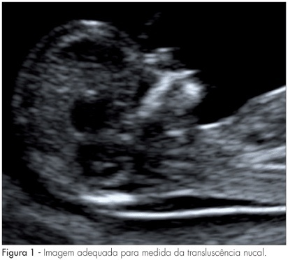
We have found that gestational age has correlation with ductus venosus, nuchal translucency, free fraction of β subunit of human chorionic gonadotropin, pregnancy-associated plasma protein-A and placental growth factor; there is also a correlation between history of miscarriages and nasal bone. Furthermore, we correlated body mass index with nuchal translucency, free fraction of β subunit of human chorionic gonadotropin, and pregnancy-associated plasma protein-A. Maternal age was associated with free fraction of β subunit of human chorionic gonadotropin and pregnancy-associated plasma protein-A.
Our study demonstrates for the first time the behavior of the biochemical and ultrasonographic markers of chromosomopathy screenings during the first trimester in euploid fetuses in Colombia. Our information is consistent with international reference values. Moreover, we have shown the correlation of different variables with maternal characteristics to determine the variables that could help with development of a screening process during the first trimester with high detection rates.
Summary
Revista Brasileira de Ginecologia e Obstetrícia. 2022;44(7):646-653
This study aims to describe the behavior of chromosomopathy screenings in euploid fetuses.
This is a prospective descriptive study with 566 patients at 11 to 14 weeks of gestation. The associations between ultrasound scans and serological variables were studied. For the quantitative variables we used the Spearman test; for the qualitative with quantitative variables the of Mann-Whitney U-test; and for qualitative variables, the X2 test was applied. Significance was set at p ≤ 0.05.
We have found that gestational age has correlation with ductus venosus, nuchal translucency, free fraction of β subunit of human chorionic gonadotropin, pregnancy-associated plasma protein-A and placental growth factor; there is also a correlation between history of miscarriages and nasal bone. Furthermore, we correlated body mass index with nuchal translucency, free fraction of β subunit of human chorionic gonadotropin, and pregnancy-associated plasma protein-A. Maternal age was associated with free fraction of β subunit of human chorionic gonadotropin and pregnancy-associated plasma protein-A.
Our study demonstrates for the first time the behavior of the biochemical and ultrasonographic markers of chromosomopathy screenings during the first trimester in euploid fetuses in Colombia. Our information is consistent with international reference values. Moreover, we have shown the correlation of different variables with maternal characteristics to determine the variables that could help with development of a screening process during the first trimester with high detection rates.
Summary
Revista Brasileira de Ginecologia e Obstetrícia. 2017;39(12):647-652
To determine cervical biometry in pregnant women between 18 and 24 weeks of gestation and the ideal mode of measurement of cervical length in cases of curved and straight cervical morphology.
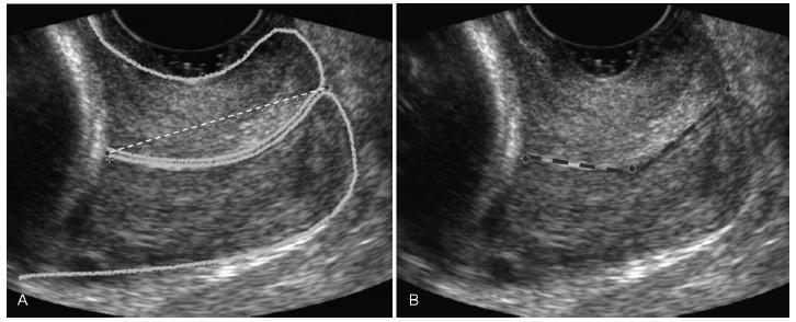
The uterine cervices of 752 low-risk pregnant women were assessed using transvaginal ultrasound in a prospective cross-sectional study. In women with straight uterine cervices, cervical biometry was performed in a continuous manner. In women with curved uterine cervices, the biometry was performed using both the continuous and segmented techniques (in segments joining the cervical os). Polynomial regression models were created to assess the correlation between the cervical length and gestational age. The paired Student t-test was used to comparemeasuring techniques.
The cervical biometry results did not vary significantly with the gestational age and were best represented by linear regression (R2 = 0.0075 with the continuous technique, and R2 = 0.0017 with the segmented technique). Up to the 21st week of gestation, there was a predominance of curved uterine cervix morphology (58.9%), whereas the straight morphology predominated after this gestational age (54.2%). There was a significant difference between the continuous and the segmented measuring methods in all the assessed gestational ages (p < 0.001).
Cervical biometry in pregnant women between 18 and 24 weeks was represented by a linear regression, independently of the measuring mode. The ideal measuring technique was the transvaginal ultrasound performed at a gestational age 21 weeks.
Summary
Revista Brasileira de Ginecologia e Obstetrícia. 2017;39(12):647-652
To determine cervical biometry in pregnant women between 18 and 24 weeks of gestation and the ideal mode of measurement of cervical length in cases of curved and straight cervical morphology.
The uterine cervices of 752 low-risk pregnant women were assessed using transvaginal ultrasound in a prospective cross-sectional study. In women with straight uterine cervices, cervical biometry was performed in a continuous manner. In women with curved uterine cervices, the biometry was performed using both the continuous and segmented techniques (in segments joining the cervical os). Polynomial regression models were created to assess the correlation between the cervical length and gestational age. The paired Student t-test was used to comparemeasuring techniques.
The cervical biometry results did not vary significantly with the gestational age and were best represented by linear regression (R2 = 0.0075 with the continuous technique, and R2 = 0.0017 with the segmented technique). Up to the 21st week of gestation, there was a predominance of curved uterine cervix morphology (58.9%), whereas the straight morphology predominated after this gestational age (54.2%). There was a significant difference between the continuous and the segmented measuring methods in all the assessed gestational ages (p < 0.001).
Cervical biometry in pregnant women between 18 and 24 weeks was represented by a linear regression, independently of the measuring mode. The ideal measuring technique was the transvaginal ultrasound performed at a gestational age 21 weeks.
Summary
Revista Brasileira de Ginecologia e Obstetrícia. 2007;29(12):647-653
DOI 10.1590/S0100-72032007001200008
Screening for major chromosomal abnormalities can be provided in the first trimester of pregnancy. Screening by a combination of fetal nuchal translucency and maternal serum free human chorionic gonadotropin and pregnancy-associated plasma protein-A can identify 90% of fetuses with trisomy 21 and other major chromosomal abnormalities for a false-positive rate of 5%. This is superior to the 30% detection rate achieved by maternal age and 65% by second-trimester maternal serum biochemistry. A further improvement in the effectiveness of first-trimester screening is likely to be achieved by a risk-orientated two-stage approach. In this approach, the patients are subdivided into a high-risk group, requiring invasive testing; a low-risk group, which can be reassured that an abnormality is unlikely, and an intermediate-risk group (risk of 1 in 101 to 1 in 1000), in which further assessment is performed by first-trimester ultrasound examination (for presence/absence of the nasal bone or presence/absence of tricuspid regurgitation or normal/abnormal Doppler velocity waveform in the ductus venosus), and chorionic villus sampling is performed if their adjusted risk becomes 1 in 100 or more. Those performing first-trimester scans should be appropriately trained and their results subjected to external quality assurance. This process was well established by the Fetal Medical Foundation several years ago and is widely accepted internationally.